A 44-year-old comes in with eight months of right-sided neck pain. He has had a chiropractor, a massage therapist, a course of physical therapy, and an MRI report listing bulging discs at C5–6 and C6–7 with “facet arthropathy,” which is what most 44-year-old necks look like on MRI. Nobody has examined his arm. Nobody has pressed on the joints beside his spine. Nobody has asked whether his head drifts. The ache is real, and the reason nothing has fixed it is that nobody has yet identified which of four different structures is producing it.
That is the visit at Modal Pain Management. Dr. Alex Movshis, dual board-certified in Anesthesiology and Pain Medicine, examines the neck, the arm, and the head posture, reads any imaging against the exam, and names the most likely source — a pinched nerve, a facet joint, cervical dystonia, or myofascial trigger points — because each one gets a different procedure. This page walks through the four, the headache that comes from the neck, the red flags, what the MRI can and cannot tell you, and what to expect at our Midtown Manhattan office at 369 Lexington Avenue.
Four necks that feel the same and need four different procedures
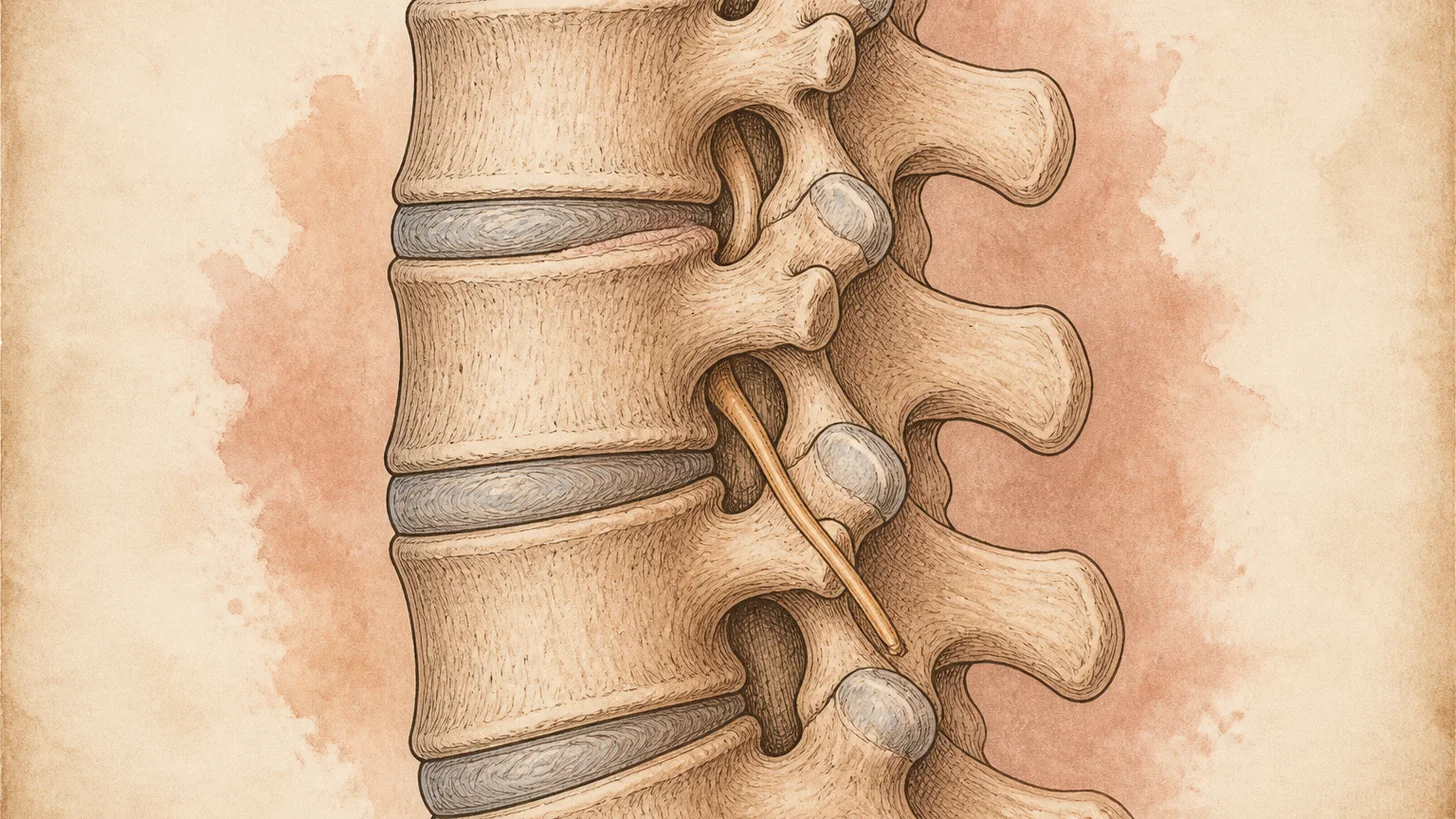
A pinched nerve (cervical radiculopathy). A disc herniation or a bone spur at C5–6 or C6–7 irritates the nerve root leaving the spine, and the pain travels: down the outer arm to the thumb and index finger for C6, down the back of the arm to the middle finger for C7, along the inner forearm to the ring and small fingers for C8. Tingling or numbness follows the same strip, and weakness — bending the elbow, extending the wrist, straightening the fingers, gripping — names the level. Looking up and turning toward the painful side often reproduces it (the Spurling maneuver), and resting the hand on top of the head can relieve it. Most cases settle over weeks to months. The ones that do not, or that come with weakness, are treated with a fluoroscopically guided cervical epidural steroid injection at the level the exam and the MRI agree on. Our guide to a pinched nerve in the neck covers the levels, the timeline, and when surgery enters the picture.
A facet joint. The small paired joints at the back of each cervical level are a common source of chronic neck pain after a car accident, and of neck pain without any injury. The pain is deep and one-sided, worse leaning the head back or turning toward the painful side, and tender directly over the joint a finger’s breadth from the midline. It refers in a map worked out by injecting the joints of 61 patients and reproducing their usual pain: C2–3 to the back of the head, C3–4 and C4–5 to the back of the neck, C4–5 and C5–6 to the top of the shoulder, and C6–7 and C7–T1 toward the shoulder blade (Fukui 1996). There are no arm symptoms and no weakness. The diagnosis cannot be made on MRI, because facet arthritis is common in necks over 40. A medial branch block tests it, and in people whose pain clearly responds, the next step is radiofrequency ablation of the same nerves. In the sham-controlled trial that established the procedure, 24 patients with neck pain after whiplash, selected by controlled blocks, relief after radiofrequency neurotomy lasted a median 263 days, against 8 days after sham (Lord 1996). Not everyone responds, and the relief fades over time in those who do.
Cervical dystonia. The head turns, tilts, or pulls on its own, the muscles on the pulling side are thick and sore, and a light touch to the chin or cheek quiets the pull — the sensory trick that patients rarely mention and clinicians rarely ask about. It is a neurological movement disorder, not a spine problem, and it is routinely treated as a stiff neck for years: in a survey of 108 patients at a U.S. academic center, people saw an average of 3.5 providers over about 44 months before diagnosis (Tiderington 2013). The treatment is EMG-guided botulinum toxin, with ultrasound for the deep muscles, into the specific overactive muscles, repeated about every 12 weeks. The full picture — the head positions, the tremor, the diagnosis, the injections — is on our cervical dystonia page.
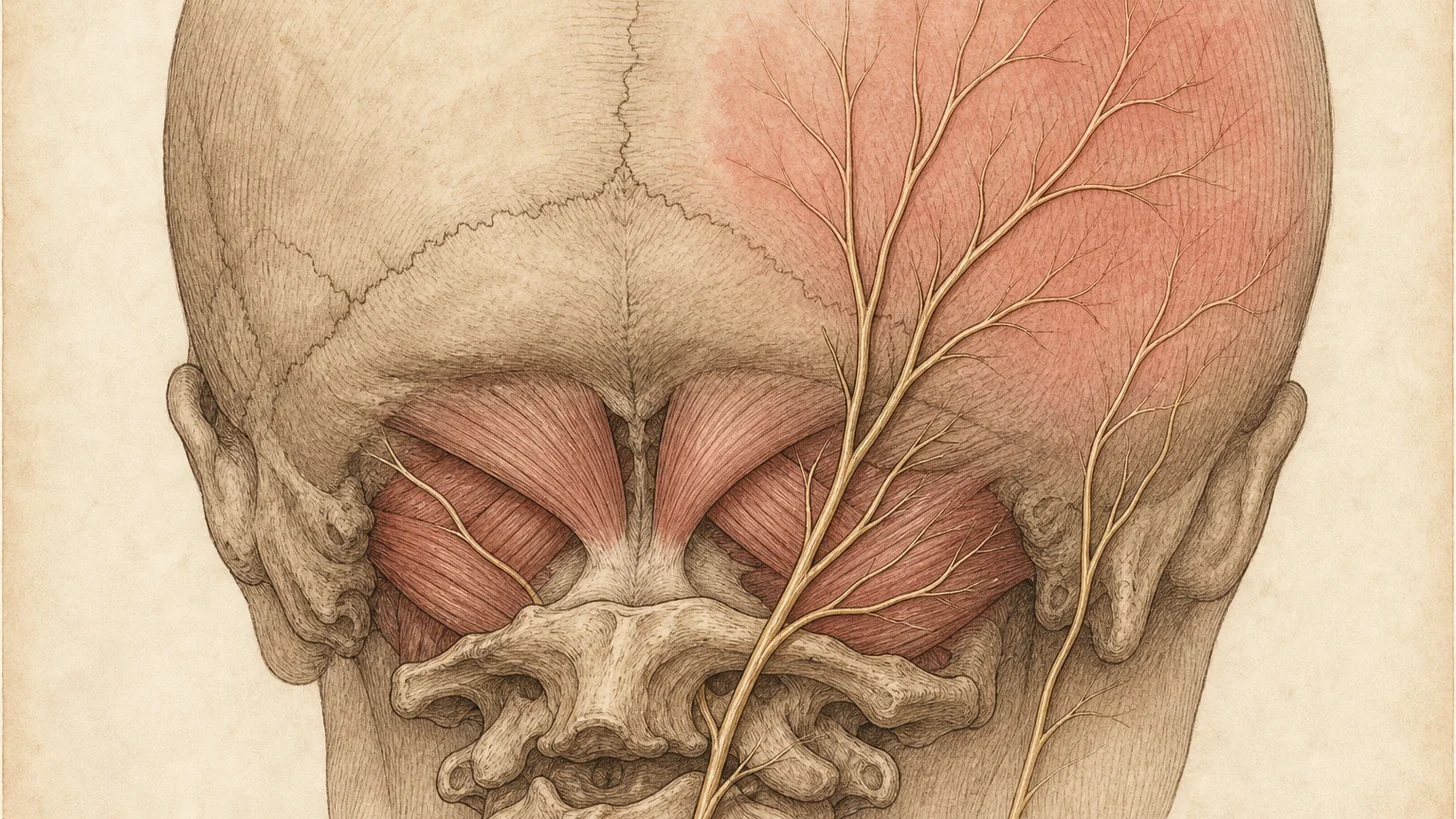
Myofascial neck pain. The trapezius and the levator scapulae, held short for hours over a screen, develop taut bands with trigger points that reproduce the patient’s pain when pressed and refer a dull ache toward the shoulder blade or the back of the head. The neck moves fully, the arm examines normally, and the pain tracks the workday. Posture and loading changes are the treatment for most patients, and our guide to tech neck covers them. For the trigger points that do not release, trigger point injections can calm them so the program can work. Myofascial pain also sits on top of the other three — a facet joint or a dystonic pull keeps the muscles around it guarded — which is why it is treated alongside the primary source rather than instead of it.
The red flags that skip the sorting
Clumsy hands, difficulty with buttons or handwriting, an unsteady walk, or hyperactive reflexes point to the spinal cord itself being compressed (cervical myelopathy), which is a surgical question, not an injection question, and we refer the same week. If these come on suddenly or worsen quickly, call 911 or go to the nearest emergency room. Neck pain after significant trauma needs emergency imaging before anyone moves the neck: call 911. Fever with neck pain, or neck pain in a patient with cancer, intravenous drug use, or a weakened immune system, needs same-day medical evaluation to rule out infection or tumor. A sudden severe headache with neck pain, especially with dizziness, double vision, or slurred speech, can be a tear in the vertebral artery. Call 911 or go to the nearest emergency room. We see these rarely and send them to the right place the same day.
What the MRI can and cannot tell you
Cervical MRI is the right test when the exam suggests a pinched nerve and an epidural injection or surgery is being planned, when there are signs of cord compression, and after trauma. It is a poor test for “what hurts” on its own. In 1,211 people with no neck symptoms, 87.6 percent had disc bulging on cervical MRI and 5.3 percent had some spinal cord compression they did not know about (Nakashima 2015). The report that lists bulging discs at three levels and facet arthropathy has described a common 44-year-old neck, not identified a pain generator. The exam narrows that down, and a diagnostic injection, a medial branch block for the joint or a selective nerve root block when the level is in doubt, adds evidence. If you already have an MRI, bring it. It is read against the exam.
The procedures, and what each one is for
The cervical epidural steroid injection is for the inflamed nerve root. It is done under fluoroscopy, with contrast to check the needle position, following the safeguards a multidisciplinary working group and national societies agreed on (Rathmell 2015). It can ease the painful phase of a radiculopathy while most roots settle on their own. It is not a treatment for axial neck pain without arm symptoms, and it is not repeated indefinitely.
The medial branch block and radiofrequency ablation are for the facet joint. The block is the test. In people who respond, ablation of the same medial branches can give months of relief. Relief can fade as the treated nerve recovers, and the procedure can be repeated.
Botulinum toxin, Botox™, Dysport™, or Daxxify™, is for cervical dystonia, injected under EMG guidance, with ultrasound for the deep muscles, into the specific muscles that are pulling.
Trigger point injections are for the taut bands in the trapezius and levator scapulae, placed by feel or under ultrasound depending on the muscle. The third occipital nerve block and the occipital nerve block are for the headaches that come from the neck. Physical therapy runs alongside every one of these, and the neck pain exercises and treatments guide covers the program for the muscular component. What we do not do is surgery: a nerve root that has failed injections with persistent weakness, a myelopathy, or an unstable segment goes to a spine surgeon with the exam and the block results attached, and that referral is part of the visit rather than a failure of it.
What to expect at Modal Pain Management
The first visit takes 45 to 60 minutes and may include an ultrasound exam. Bring any imaging, the list of what has been tried, and — if your head drifts or a touch to the face eases the pull — say so, because it changes the diagnosis. Dr. Movshis examines the neck, the arm, and the head posture, reads the imaging against the exam, and names the most likely source and the procedure that tests or treats it. Procedures are typically scheduled within 1–2 weeks at the same office, take 15–30 minutes, and you go home the same day. Follow-up measures the response and either holds the plan, escalates to ablation or a different target, or goes to a surgical opinion. The consultation and the procedures on this page are covered by most commercial PPO plans, some with prior authorization that our office handles. The practice does not accept Medicare, Medicaid, HMO plans, or workers’ compensation. Self-pay prices are published per procedure, and you can verify your insurance before the visit.
If your neck pain has had a chiropractor, a massage, a physical therapy course, and an MRI, and nobody has yet said which structure it is, that is the visit to book.
References
- Fukui S, Ohseto K, Shiotani M, et al. Referred pain distribution of the cervical zygapophyseal joints and cervical dorsal rami. Pain. 1996;68(1):79–83. doi:10.1016/S0304-3959(96)03173-9 · PubMed
- Lord SM, Barnsley L, Wallis BJ, McDonald GJ, Bogduk N. Percutaneous radio-frequency neurotomy for chronic cervical zygapophyseal-joint pain. N Engl J Med. 1996;335(23):1721–1726. doi:10.1056/NEJM199612053352302 · PubMed
- Tiderington E, Goodman EM, Rosen AR, et al. How long does it take to diagnose cervical dystonia? J Neurol Sci. 2013;335(1–2):72–74. doi:10.1016/j.jns.2013.08.028 · PubMed
- Nakashima H, Yukawa Y, Suda K, Yamagata M, Ueta T, Kato F. Abnormal findings on magnetic resonance images of the cervical spines in 1211 asymptomatic subjects. Spine. 2015;40(6):392–398. doi:10.1097/BRS.0000000000000775 · PubMed
- Rathmell JP, Benzon HT, Dreyfuss P, et al. Safeguards to prevent neurologic complications after epidural steroid injections: consensus opinions from a multidisciplinary working group and national organizations. Anesthesiology. 2015;122(5):974–984. doi:10.1097/ALN.0000000000000614 · PubMed