Knee bursitis is one of the most common — and most under-treated — sources of knee pain in adults. At Modal Pain Management in Midtown Manhattan, Dr. Alex Movshis uses real-time ultrasound to identify the specific bursa involved, drain any fluid collection, and deliver targeted anti-inflammatory medication directly into the bursa — typically all in a single office visit. Most patients walk out the same day with substantially less pain, return to work the next morning, and resume normal activities within one to two weeks.
This page covers what knee bursitis is, the five distinct types (each with its own pain pattern, causes, and treatment), how it is diagnosed at Modal Pain Management, and the evidence-based treatment ladder we use to get patients back to walking, running, kneeling, and climbing stairs without pain.
What Is a Bursa, and What Is Knee Bursitis?
A bursa is a small, flat fluid-filled sac that sits between bone and overlying soft tissue (skin, tendon, or muscle) at points of friction. The knee has roughly a dozen named bursae. Each one is normally a thin film of synovial fluid that lubricates motion and protects the underlying tissue from pressure. When a bursa becomes irritated — through repetitive pressure, direct trauma, overuse, infection, or systemic inflammation — it fills with fluid, swells, and becomes painful. That inflamed-and-distended state is bursitis.
The five clinically important knee bursae are:
- Prepatellar bursa — sits superficially over the front of the kneecap; inflamed by direct kneeling pressure.
- Infrapatellar bursae (superficial and deep) — sit just below the kneecap; inflamed by jumping, running, and patellar tendon overuse.
- Pes anserine bursa — sits on the inner shin about 2 inches below the joint line where three tendons (sartorius, gracilis, semitendinosus) insert; inflamed by overuse and often coexists with medial knee osteoarthritis.
- Suprapatellar bursa — communicates with the joint itself in most adults; inflammation here usually reflects a knee joint problem (osteoarthritis, meniscus tear, synovitis).
- Semimembranosus bursa — sits behind the knee; when distended it forms a Baker’s cyst.
The treatment depends entirely on which bursa is involved, what is driving the inflammation, and whether infection is present. Generic “knee bursitis” advice — ice, rest, ibuprofen — is reasonable as a first step but misses the diagnostic specificity that determines outcomes.
The Five Types of Knee Bursitis
1. Prepatellar Bursitis (“Housemaid’s Knee,” “Carpet Layer’s Knee”)
Prepatellar bursitis is the most common knee bursitis and the easiest to diagnose: a soft, squishy, often golf-ball-sized swelling sits directly over the front of the kneecap. The skin may be tender to touch and painful when you kneel, but the knee joint itself usually bends and straightens normally. Historically named for occupations involving prolonged kneeling — housemaids, clergymen, carpet layers, plumbers, gardeners, roofers — it remains an occupational diagnosis today.
Acute traumatic prepatellar bursitis (a fall on the knee) often resolves with ice, NSAIDs, and a knee pad in 1–3 weeks. Chronic, occupation-driven prepatellar bursitis usually requires aspiration plus corticosteroid injection plus permanent kneeling-pad use to prevent recurrence. Septic prepatellar bursitis — which presents with warmth, redness, fever, and often a small abrasion or insect bite over the kneecap — must be aspirated for culture before any steroid is given.
2. Infrapatellar Bursitis (“Clergyman’s Knee,” “Jumper’s Knee Bursitis”)
There are two infrapatellar bursae: a superficial one between the patellar tendon and skin, and a deep one between the patellar tendon and the tibia. Inflammation causes burning or aching pain just below the kneecap, worsened by jumping, running downhill, prolonged kneeling in an upright posture, or kicking. It is most common in basketball players, volleyball players, runners, and dancers. Infrapatellar bursitis frequently coexists with patellar tendinopathy (“jumper’s knee”) — distinguishing them requires ultrasound, which Modal Pain Management performs at the consultation visit.
3. Pes Anserine Bursitis (Inner Knee Pain)
Pes anserine bursitis causes a deep, aching pain on the inner side of the knee, about 2 inches below the joint line. It is common in middle-aged and older adults, especially women, and frequently coexists with medial compartment osteoarthritis. Patients typically report pain when climbing stairs, rising from a chair, or sleeping with their knees touching at night. Risk factors include obesity, valgus (knock-knee) alignment, weak hip abductors, and tight hamstrings. Treatment combines ultrasound-guided corticosteroid injection with a structured physical therapy program targeting hip strength and quadriceps balance — addressing the bursitis without addressing the biomechanics leads to predictable recurrence.
4. Suprapatellar Bursitis
The suprapatellar bursa communicates with the knee joint itself in most adults, so inflammation here usually reflects an intra-articular problem: knee osteoarthritis, a meniscus tear, inflammatory arthritis (rheumatoid, psoriatic, gout), or post-traumatic synovitis. The clinical presentation is a fluid-filled fullness above the kneecap. Treatment is directed at the underlying joint pathology — intra-articular corticosteroid or hyaluronic acid injection, physical therapy, and weight management — rather than at the bursa in isolation.
5. Semimembranosus Bursitis (Baker’s Cyst)
A Baker’s cyst is a distended semimembranosus bursa in the popliteal fossa (behind the knee). Most adult Baker’s cysts in middle-aged and older patients communicate with the knee joint and reflect an underlying meniscus tear or osteoarthritis. Symptoms include a tightness, fullness, or visible bulge behind the knee that worsens with prolonged standing or full knee flexion. A ruptured Baker’s cyst can mimic deep vein thrombosis (calf pain, swelling, warmth) — distinguishing them requires urgent ultrasound. Treatment focuses on the underlying joint problem; aspiration with steroid is sometimes performed for symptomatic large cysts.
How Knee Bursitis Is Diagnosed at Modal Pain Management
Diagnosis at Modal Pain Management is anatomically specific from the first visit. Dr. Movshis performs a focused history (occupation, kneeling exposure, sports, trauma, prior knee surgery, systemic conditions like gout or rheumatoid arthritis), a knee exam locating tenderness over the specific bursa, and bedside musculoskeletal ultrasound during the consultation itself. Ultrasound directly visualizes:
- The bursa fluid collection — confirming the diagnosis and which specific bursa is inflamed.
- The bursa wall — distinguishing simple bursitis from chronic synovial thickening or septic bursitis.
- Adjacent structures — patellar tendon (rule out tendinopathy or tear), medial meniscus (rule out coexisting tear in pes anserine cases), popliteal vessels (rule out aneurysm or DVT in suspected Baker’s cyst), and the knee joint itself (rule out an effusion that needs separate management).
If the bursa is large or the clinical picture suggests possible infection, fluid is aspirated and sent to the lab for cell count, Gram stain, culture, and crystal analysis. X-rays are added when osteoarthritis is suspected. MRI is reserved for atypical, complex, or surgical cases.
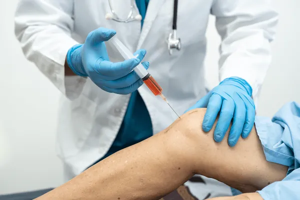
Image-Guided Treatment of Knee Bursitis
The cornerstone of treatment for symptomatic, fluid-filled knee bursitis is ultrasound-guided aspiration with corticosteroid injection. Performed in a 15-minute office procedure, the technique involves:
- Skin preparation and local anesthesia — the skin over the bursa is sterilized and numbed with lidocaine.
- Ultrasound-guided needle placement — a needle is advanced under real-time ultrasound directly into the bursa, avoiding the patellar tendon, popliteal vessels, and joint capsule.
- Aspiration — fluid is withdrawn and inspected; sent for laboratory analysis if infection or crystal disease is suspected.
- Corticosteroid injection — once aspiration is complete (and infection is ruled out), a small volume of corticosteroid mixed with local anesthetic is deposited into the now-decompressed bursa.
- Compressive dressing and discharge — a compression sleeve is applied; patients walk out the same day.
Image guidance matters. Blind (landmark-based) bursa injections miss the bursa in 30–40% of cases, leading to underwhelming results that often get attributed to “bursitis just being a hard problem to treat” when in fact the medication never reached the target. Ultrasound guidance pushes accuracy to 95%+ and is the standard of care for every bursa procedure at Modal Pain Management.
For chronic, recurrent knee bursitis that has failed two appropriately performed image-guided injections, image-guided genicular nerve radiofrequency ablation is offered as a non-surgical option that can interrupt the pain pathway for 6–12 months at a time.
Physical Therapy and Self-Care
Injection alone is insufficient when biomechanical factors are driving the inflammation. Patients with pes anserine bursitis, infrapatellar bursitis, or recurrent prepatellar bursitis benefit from a structured course of physical therapy targeting:
- Quadriceps strength — particularly vastus medialis activation to protect the patellofemoral joint.
- Hip abductor and external rotator strength — to control valgus knee collapse during gait and stair climbing.
- Hamstring and calf flexibility — reducing posterior knee tension that worsens infrapatellar and Baker’s cyst symptoms.
- Gait retraining — for runners, addressing overstriding, cadence, and shoe wear.
- Kneeling-load reduction — for occupational cases, replacing direct kneeling with kneeling pads, foam mats, or alternate work positions.
Home care includes ice 15–20 minutes 3–4 times daily during flares, NSAIDs (naproxen 220–440 mg twice daily, ibuprofen 400–600 mg three times daily) for 7–14 days unless contraindicated, a compressive knee sleeve during activity, and avoidance of kneeling, deep squats, and high-impact loading until symptoms have resolved for at least two weeks.
When Knee Bursitis Is an Emergency: Septic Bursitis
Septic bursitis — bacterial infection of the bursa — is a medical emergency. Untreated, the infection can spread to the joint itself, the bone, or the bloodstream. Warning signs are warmth, redness, marked tenderness, fever, chills, and often a small cut, scrape, abrasion, insect bite, or recent injection over the bursa. Diabetic patients, immunosuppressed patients, and patients on long-term corticosteroids are at higher risk.
If septic bursitis is suspected, fluid must be aspirated immediately and sent for Gram stain and culture. Empirical antibiotics covering Staphylococcus aureus (the most common organism) are started while culture results are pending. Corticosteroid is never injected into a potentially infected bursa. Most cases respond to outpatient antibiotics; severe or treatment-refractory cases may require IV antibiotics or surgical drainage.
If you have any concern that your knee bursitis may be infected — particularly fever combined with knee swelling and redness — go to an emergency room or urgent care the same day rather than waiting for an outpatient appointment.
Why Modal Pain Management for Knee Bursitis in NYC
Modal Pain Management is a physician-led interventional pain practice located at 369 Lexington Avenue, Floor 25 in Midtown Manhattan (NYC 10017), led by Dr. Alex Movshis, a dual board-certified pain management physician fellowship-trained at the Icahn School of Medicine at Mount Sinai. Every patient is evaluated and treated by Dr. Movshis personally — not by a rotating cast of providers. Bedside musculoskeletal ultrasound is available at the consultation visit, which means most knee bursitis cases are diagnosed and treated in a single appointment. Same-week new-patient appointments are typically available.
For sourced clinical evidence on the procedures referenced on this page — including efficacy ranges, recovery timelines, and comparison data — see the clinical evidence and citations page. Most commercial PPO insurance plans are accepted; Medicare and Medicaid are not. Insurance benefits are verified before your visit at no charge or obligation through the insurance verification form.