Peripheral nerve entrapments share a frustrating property: they are mostly invisible on standard imaging, they reproduce clinically with bedside maneuvers that most practices do not perform, and they are most reliably diagnosed by an image-guided peripheral nerve block — a diagnostic tool that is not routinely offered outside dedicated interventional pain practices. The result is that patients with classic entrapment patterns are routinely worked up for years against the wrong differential, accumulating MRIs that look “unremarkable” while the right diagnosis takes 15 minutes at the bedside.
The diagnostic block is the workhorse. A small volume of local anesthetic delivered onto or immediately adjacent to the suspected nerve produces a clean diagnostic answer in 10–15 minutes. Image guidance is essential — blind landmark technique runs accuracy in the 50–70% range for these nerves; ultrasound and fluoroscopy push accuracy above 95%. At Modal Pain Management, 369 Lexington Avenue Floor 25 in Midtown Manhattan, Dr. Alex Movshis (dual board-certified in Anesthesiology and Pain Medicine, fellowship-trained at the Icahn School of Medicine at Mount Sinai) performs the full range of image-guided peripheral nerve blocks and coordinates surgical referral when conservative care reaches its ceiling.
Common entrapments we evaluate
Meralgia paresthetica — lateral femoral cutaneous at the inguinal ligament
The pattern is anchored by PAT-005 above. The classic non-GLP-1 triggers are unchanged: tight waistbands, low-rise jeans, tactical or weightlifting belts, prolonged hip extension (long-distance cycling, military or police duty positioning), pregnancy, diabetes, post-surgical iatrogenic injury (anterior approach total hip arthroplasty, iliac crest bone graft, inguinal hernia repair), and central obesity. The diagnostic and therapeutic workup is the same regardless of trigger: ultrasound-guided LFCN block first (diagnostic in 15 minutes, therapeutic 6–12 weeks with steroid), hydrodissection second for refractory cases, pulsed RFA third for chronic disease, and peripheral nerve surgical decompression or PNS as salvage. Detailed coverage on the dedicated meralgia paresthetica page.
Anterior cutaneous nerve entrapment syndrome (ACNES)
Cluneal nerve entrapment — superior, middle, inferior
Wartenberg’s syndrome — superficial radial at the wrist
Pronator and anterior interosseous syndromes
The median nerve has two well-described entrapment sites in the forearm distinct from carpal tunnel at the wrist. Pronator teres syndrome is median nerve compression as it passes between the two heads of the pronator teres muscle in the proximal forearm. Pain is in the volar forearm; numbness involves the median-innervated digits (thumb, index, middle, radial half of ring) but the thenar muscles are spared because the recurrent motor branch arises distal to the wrist. The diagnostic move is reproducing the pain with resisted pronation. Anterior interosseous syndrome is compression of the AIN — the pure motor branch of the median nerve in the proximal forearm. It produces weakness in flexor pollicis longus, flexor digitorum profundus to index and middle, and pronator quadratus — the “OK sign” is lost (the thumb-index pinch cannot make a circle). Both syndromes are diagnosed by EMG/NCS plus ultrasound-guided diagnostic block at the entrapment site, and refractory cases are referred to hand surgery for decompression.
Quadrilateral space syndrome — axillary nerve posteriorly
The axillary nerve and posterior humeral circumflex artery pass through the quadrilateral space (bounded by teres minor, teres major, long head of triceps, and humerus). Compression in this space — typically from posterior shoulder overuse (volleyball, baseball, swimming), fibrous bands, or labral cyst — produces posterior shoulder pain and weakness of the deltoid and teres minor. Patients describe pain along the lateral upper arm with overhead activity. Diagnosis combines clinical exam (positive test with abduction + external rotation reproducing the pain), high-resolution ultrasound or MRI of the quadrilateral space, and image-guided diagnostic axillary nerve block.
Suprascapular nerve at the spinoglenoid notch
The suprascapular nerve passes under the transverse scapular ligament at the suprascapular notch and around the base of the scapular spine at the spinoglenoid notch. Either point is an entrapment site — most commonly at the spinoglenoid notch from a paralabral cyst (associated with posterior labral tear), and at the suprascapular notch from repetitive overhead activity or post-surgical scar. The presentation is deep posterior shoulder pain with infraspinatus weakness (external rotation), often with teres minor wasting visible on inspection in chronic cases. Diagnostic ultrasound-guided suprascapular nerve block at the notch is both diagnostic and therapeutic for 6–12 weeks. Refractory cases are referred to shoulder surgery for decompression and labral repair when a paralabral cyst is the driver.
Tarsal tunnel syndrome — posterior tibial at the medial malleolus
The tibial nerve passes behind the medial malleolus through the tarsal tunnel — the foot’s anatomic analog of carpal tunnel. Compression at this point produces burning, electric pain in the sole of the foot, often radiating into the toes, worse with prolonged standing and improved with rest. Tarsal tunnel is dramatically under-recognized and is frequently misdiagnosed as plantar fasciitis (which has a different pain pattern — first-step morning pain, focal at the medial calcaneal tubercle, no burning component). Diagnostic ultrasound-guided posterior tibial nerve block at the tarsal tunnel confirms the diagnosis. Surgical referral to foot and ankle surgery for tarsal tunnel release is the salvage for refractory cases (Dellon-style decompression).
Common peroneal entrapment at the fibular neck
The common peroneal nerve wraps around the fibular neck immediately distal to the lateral knee, where it lies subcutaneously and is vulnerable to direct compression — tight knee-high boots, prolonged leg-crossing, post-surgical cast or brace pressure, and (most consequentially) prolonged squatting or sitting in flexion. The presentation is foot drop with sensory loss over the dorsum of the foot. Acute foot drop after a known compressive event (anesthesia positioning, prolonged squat, hospital bed-rail pressure) is the classic story. Diagnostic ultrasound at the fibular neck identifies focal nerve thickening; ultrasound-guided diagnostic block and corticosteroid injection at the entrapment point is both diagnostic and therapeutic. Refractory cases or those with established axonal injury on EMG are referred for surgical decompression by a peripheral nerve specialist.
Why entrapments get missed
Most peripheral nerve entrapments are missed for one reason: the bedside maneuvers that diagnose them — Carnett’s for ACNES, Tinel along the iliac crest for cluneal, Tinel over the brachioradialis for Wartenberg’s, FAIR test for piriformis — are not part of the routine internal medicine, family medicine, gynecology, or orthopedic physical examination. The patient gets an MRI of the suspected region instead. Standard MRI does not show most peripheral nerve entrapments; the nerve is too small for routine sequences and the entrapment is mechanical rather than structural. The result is a “normal” MRI report that closes the diagnostic loop on a misread differential.
The fix is not better imaging. The fix is the diagnostic block. Five minutes of focused history identifying the candidate nerve, 15 minutes of ultrasound-guided block, and the diagnostic ambiguity is resolved at the bedside. The Carnett’s sign for ACNES is the canonical example — a 30-second maneuver that no published imaging study can replicate.
The other reason entrapments get missed: many of them mimic conditions that are far more commonly diagnosed. Meralgia paresthetica gets called “sciatica” or “L2/L3 radiculopathy” for months before someone notices the pain does not cross the knee. ACNES gets called IBS. Cluneal entrapment gets called facet pain. Tarsal tunnel gets called plantar fasciitis. Wartenberg’s gets called carpal tunnel. In each pairing, the wrong diagnosis is far more common in absolute terms, so the workup defaults there.
How we diagnose entrapment at Modal Pain
The new-patient consultation is 45 minutes. The diagnostic workflow:
Focused history. When did the pain start? What does it feel like (burning, electric, “pins and needles”)? Where does it map? What aggravates it (clothing, position, activity)? What relieves it? What has been tried? Recent surgery in the region? Recent weight change? Specific occupational or athletic exposure?
Cutaneous mapping. The patient marks the painful area on a body diagram with a fine-tip marker. A defined territory respecting a single cutaneous nerve distribution is the strongest single diagnostic clue.
Provocative testing. Tinel along candidate entrapment points. Carnett’s for anterior abdominal wall pain. FAIR test for piriformis-pattern pain. Specific provocative maneuvers for less common entrapments.
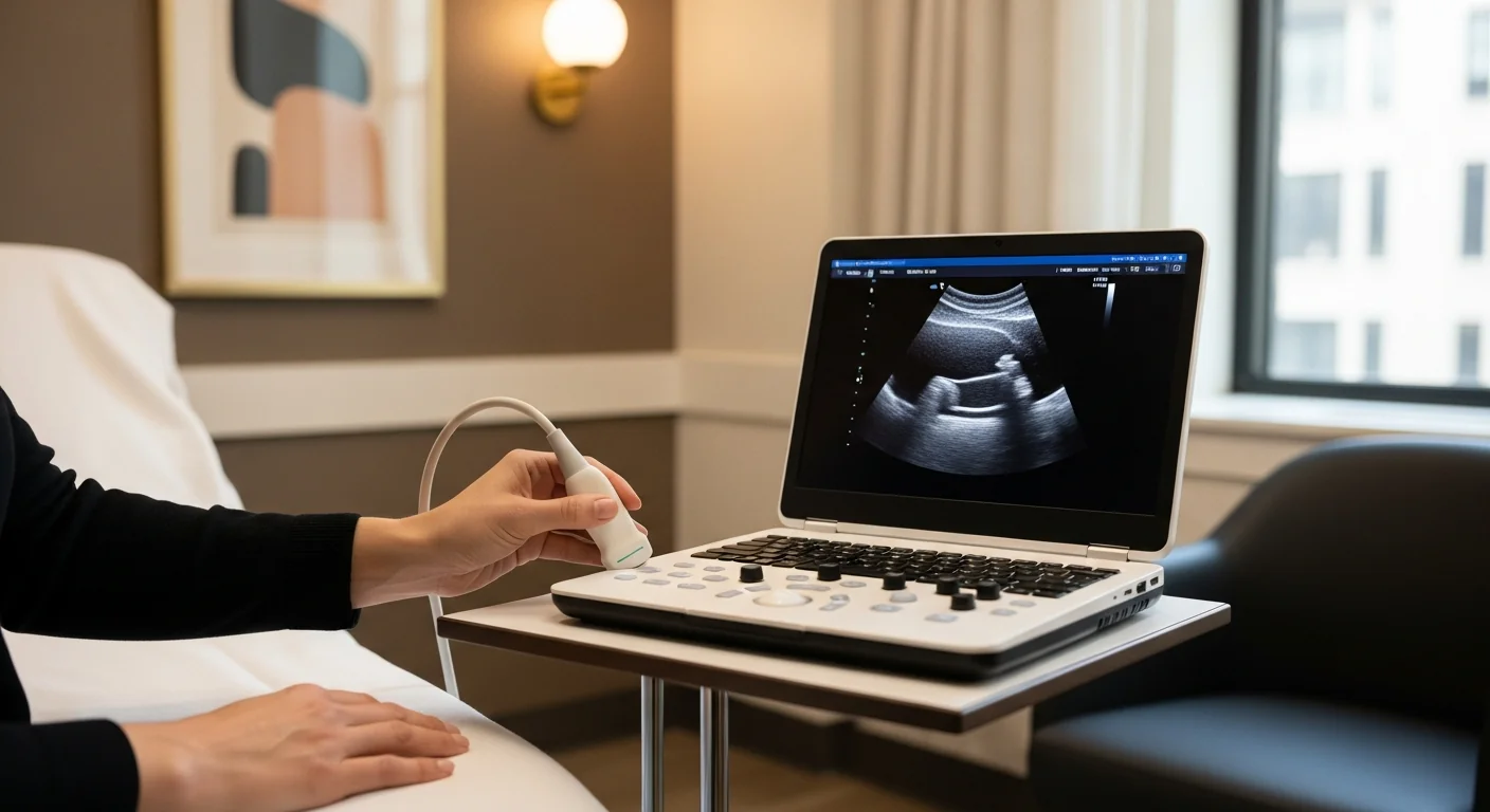
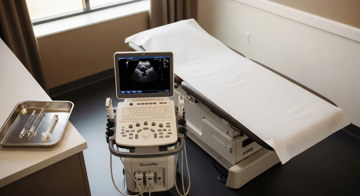
Bedside high-resolution ultrasound. Performed at the consultation. Identifies focal nerve thickening, peri-neural scar, or paralabral cyst (for suprascapular entrapment). Directly guides the diagnostic block.
Diagnostic ultrasound-guided peripheral nerve block. Performed in the same procedure suite. ≥50% pain abolition in the working window is positive; ≥75% is highly specific. The diagnostic answer is in hand within 30 minutes of the consultation visit beginning.
Treatment approach
The treatment ladder is broadly common across entrapments, with site-specific variations:
Step 1 — conservative care. Identify and remove the mechanical compressor when one exists (tight belts, watchbands, occupational positioning). Targeted physical therapy for entrapment-adjacent muscular dysfunction. Neuropathic-pain medication trial (gabapentin or pregabalin, ± tricyclic or SNRI, ± topical lidocaine patch). Most pregnancy- and weight-change-driven cases resolve completely at Step 1 once the trigger is removed.
Step 2 — image-guided diagnostic and therapeutic nerve block. Local anesthetic plus corticosteroid at the entrapment site under ultrasound guidance. Diagnostic in 10–15 minutes, therapeutic for 6–12 weeks. The block is safely repeatable.
Step 3 — ultrasound-guided hydrodissection. For nerves entrapped in dense perineural scar. Volume injection of saline or 5% dextrose mechanically frees the nerve from the surrounding fibrosis and restores nerve gliding.
Step 4 — pulsed radiofrequency ablation. For chronic refractory entrapment. Pulsed (not conventional thermal) RFA is neuromodulatory and non-destructive, producing 6–12 months of relief and safely repeatable.
Step 5 — peripheral nerve stimulation (PNS) referral. Percutaneous PNS at the affected nerve is a growing option for severe refractory entrapment with FDA clearance and strong published case-series evidence.
Step 6 — surgical referral by specialty category. Peripheral nerve plastic surgery for Dellon-style external neurolysis (Wartenberg’s, tarsal tunnel, cubital tunnel). Hand surgery for pronator and AIN syndromes. Foot and ankle surgery for tarsal tunnel release. Shoulder surgery for suprascapular decompression with labral repair when a paralabral cyst is the driver. General surgery (peripheral nerve subspecialist) for ACNES anterior neurectomy. Modal Pain coordinates these referrals directly when indicated and provides the documented diagnostic-block response that surgical programs require before accepting a referral.
Frequently asked questions
Six questions tuned to actual patient queries we receive on entrapment evaluation are rendered below; the page route ships the FAQPage JSON-LD automatically.
Related articles
- Meralgia Paresthetica: Why Your Outer Thigh Burns and What Actually Helps
- ACNES: When Years of GI Workups Miss the Real Cause of Your Abdominal Pain
- Wartenberg’s Syndrome: Forearm and Thumb Pain After a Wrist Injury
- Cluneal Nerve Entrapment: When “Low Back Pain” Is Actually a Nerve in Your Iliac Crest
- Notalgia Paresthetica: Causes, Symptoms, and Why the Itch Starts in Your Spine
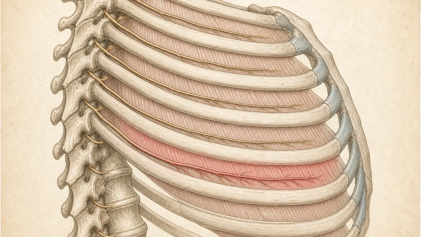
- Intercostal Neuralgia: The Band of Rib-Space Pain That Wraps Around Your Chest
- Chronic Groin Pain After Hernia Surgery: When the Nerve Got Caught
Ready to evaluate your nerve pain
If you have burning, electric, or allodynic pain in a defined cutaneous territory that has not responded to standard imaging-based workup, an image-guided diagnostic peripheral nerve block is the next step. Same-week new-patient consultations are routinely available.
Same-week diagnostic nerve block appointment Verify your insurance
Or call (646) 290-6660. Dr. Movshis sees every patient personally — initial consultation through follow-up.