If you have years of “low back and buttock pain” with negative SI joint injections, negative facet medial branch blocks, and a lumbar MRI that shows age-typical degeneration but no radicular cause for your pain — and you can press a specific point on the back of your iliac crest to reproduce the pain — you are probably reading the right post. The condition is cluneal nerve entrapment. It’s still a niche diagnosis but the literature is growing, and a 15-minute ultrasound-guided block at the iliac crest usually confirms and treats it.
Why this gets missed
The lumbosacral pain differential is a well-worn workup tree. The three branches that get explored are facet joints (treated with medial branch blocks and RFA), SI joint (treated with intra-articular injection and lateral branch RFA), and lumbar disc (treated with epidural steroid injection and physical therapy). Patients run the tree once, twice, sometimes three times across different practices. When all three come back negative or inadequate, the diagnostic options that show up next are typically psychiatric (chronic pain syndrome) or surgical (lumbar fusion for “axial low back pain”).
Neither of those is cluneal nerve entrapment. The cluneal nerves are not on the standard lumbar-spine workup tree. The patient ends up cycling through pelvic floor PT, chronic pain medication trials, and sometimes — most consequentially — a lumbar fusion that doesn’t help because it never addressed the actual pain generator.
The Maigne and Doursounian 1997 Spine paper documented 19 surgical cases. The Kuniya group has published a series of anatomic and clinical papers on middle cluneal entrapment specifically. Aota and Konno have separately validated the entrapment anatomy and characterized the “pseudo-sciatica” presentation that frequently masquerades as a lumbar radiculopathy. The body of work is small but consistent — and it confirms that cluneal entrapment is under-diagnosed because it is under-considered.
The fix is the lateral iliac-crest palpation. Palpate 7–8 cm lateral to the midline along the iliac crest with the patient prone or in a sidelying position. If a discrete tender point reproduces the patient’s typical band of buttock-and-lumbosacral pain, cluneal entrapment becomes the leading hypothesis until disproved by block.
The anatomy that matters
Three nerve groups make up the cluneal cluster:
Superior cluneal nerves — lateral cutaneous branches of the dorsal rami of L1–L3. They cross the iliac crest through a small osteofibrous tunnel formed by the thoracolumbar fascia and the bone itself, approximately 7–8 cm lateral to the midline. They then become subcutaneous and supply the skin over the upper-outer buttock. The osteofibrous tunnel is the entrapment site — anatomic variants in tunnel size, repetitive lumbar extension, prior iliac crest bone graft harvest, and idiopathic fibrotic remodeling all narrow the space.
Middle cluneal nerves — lateral cutaneous branches of the dorsal rami of S1–S3. They cross over (or sometimes through) the long posterior sacroiliac ligament and supply the central buttock skin. The Kuniya 2013 J Neurosurg Spine paper anatomically characterized entrapment at the ligament as a distinct entity from the superior cluneal entrapment at the iliac crest.
Inferior cluneal nerves — branches of the posterior femoral cutaneous nerve (S2–S3). They supply the lower buttock skin. Entrapment is less commonly diagnosed but can occur around the ischial tuberosity or under the gluteus maximus, particularly post-trauma.
All three groups are pure sensory nerves. No motor weakness, no reflex changes. The diagnostic clue is the cutaneous band reproduced by Tinel testing at the suspected entrapment point.
How we diagnose
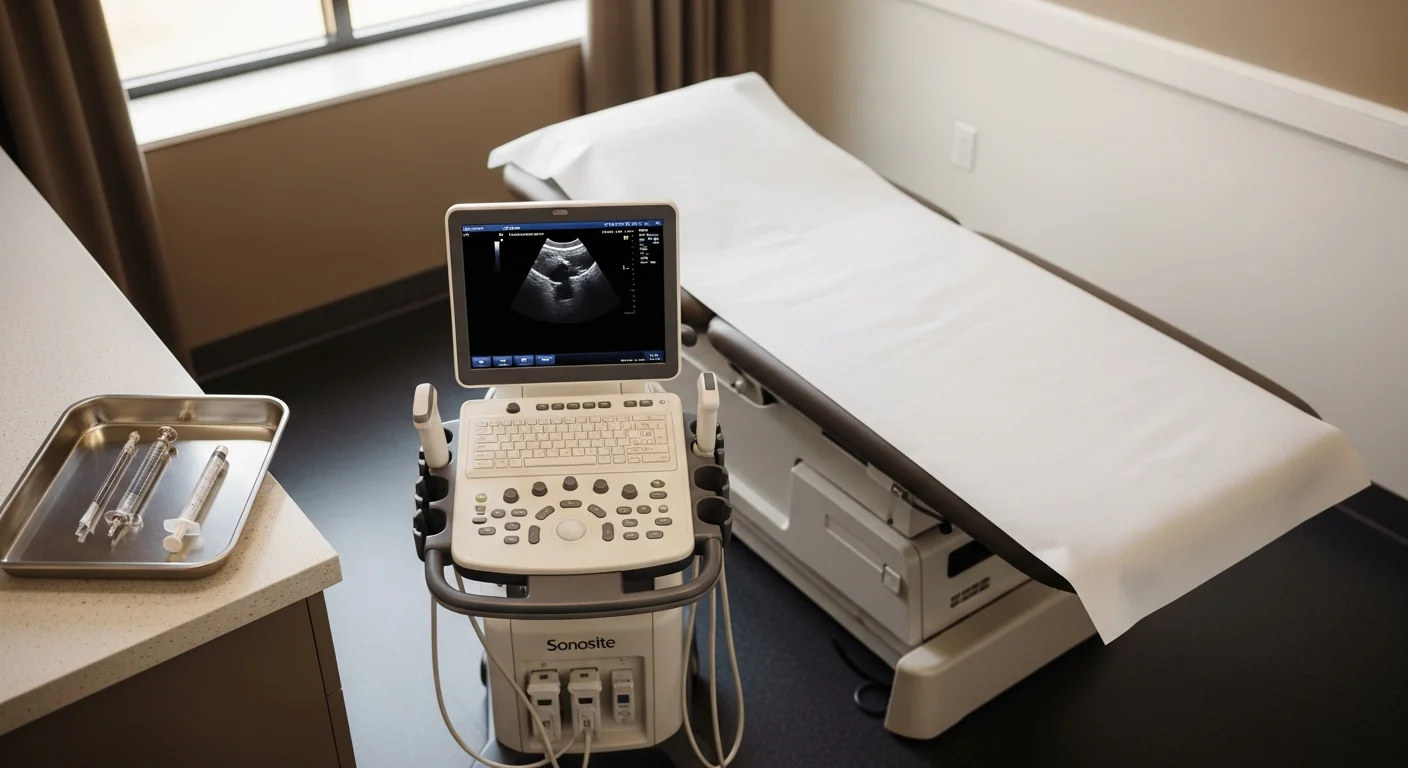
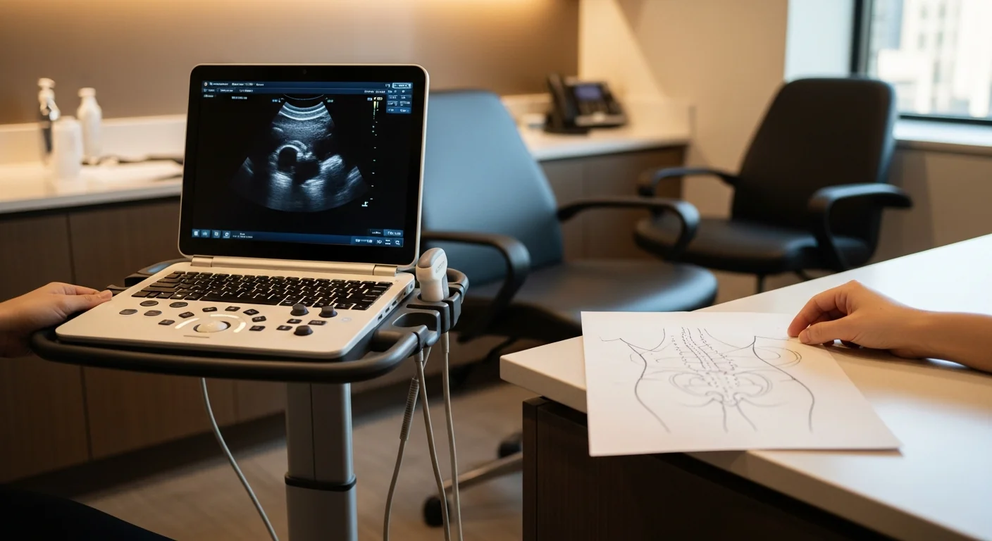
The diagnostic procedure is an ultrasound-guided cluneal nerve block at the Tinel-positive point. Performed in our procedure suite. Total visit time: about 30 minutes.
Technique for superior cluneal entrapment: with the patient in a sidelying or prone position, the posterior iliac anatomy is identified by ultrasound — the iliac crest as a bright hyperechoic line, the thoracolumbar fascia as a thin hyperechoic stripe immediately above the crest, the underlying multifidus muscle. A 22-gauge needle is advanced under direct ultrasound visualization to the fascial plane at the Tinel-positive point, typically 7–8 cm lateral to the midline. After negative aspiration, 2–3 mL of 0.5% bupivacaine plus 20 mg of triamcinolone is delivered, with spread observed in real time.
For middle cluneal entrapment: ultrasound or fluoroscopic guidance at the long posterior sacroiliac ligament, with the medication deposited in the fascial plane superficial to the ligament.
The diagnostic effect arrives within 5–10 minutes. ≥75% relief of the typical cluneal-territory pain is the definitive test. The therapeutic effect with corticosteroid added typically lasts 6–12 weeks.
Treatment ladder and when surgery is the right call
Step 1 — neuropathic-pain medication trial. Gabapentin or pregabalin at standard titration. Topical 5% lidocaine patch over the affected cutaneous band.
Step 2 — ultrasound-guided diagnostic and therapeutic cluneal nerve block. The workhorse. Diagnostic in 5–10 minutes, therapeutic for 6–12 weeks. Repeatable every 8–12 weeks.
Step 3 — ultrasound-guided hydrodissection. For nerves entrapped in dense fibrotic scar at the iliac crest tunnel — particularly common after prior iliac crest bone graft harvest or repeated steroid-only blocks that have stopped producing meaningful relief. Volume injection of saline or 5% dextrose mechanically separates the nerve from the fascial plane.
Step 4 — pulsed radiofrequency ablation at the entrapment site. For chronic refractory cases with documented positive blocks. Pulsed (not thermal) — the territory is cutaneous and the lesion must be neuromodulatory. 6–12 months of relief per treatment.
Step 5 — peripheral nerve stimulation referral. Percutaneous PNS at the cluneal nerve for severe refractory disease.
Step 6 — surgical decompression. Decompression of the cluneal nerve(s) at the entrapment site, performed by peripheral nerve plastic surgery. Maigne and Doursounian reported approximately 73% good-to-excellent outcomes; later case series have validated similar numbers. The procedure releases the osteofibrous tunnel (superior cluneal) or the long posterior sacroiliac ligament’s compressive fibers (middle cluneal). Modal Pain coordinates the referral when indicated and provides the documented diagnostic-block response surgical programs require.
The literature on cluneal entrapment is smaller than for inguinal-region neurectomies. We position surgery as the salvage option for documented refractory cases with multiple positive diagnostic blocks — not as a first-line move.
For referring physicians (especially spine and pain medicine)
When you have a chronic-low-back-pain patient with negative medial branch blocks, negative SI joint injection, and a lumbar MRI that doesn't explain the symptoms — and the cutaneous distribution stays in the buttock and lower lumbosacral region without crossing into the leg below the gluteal crease — send: the prior interventional workup summary, the lumbar MRI report, the medication trial history, and a note on whether you've palpated the iliac crest. We document a Tinel sign at the iliac crest, run an ultrasound-guided cluneal block in the room, and send notes back to your practice after each visit. For refractory cases with multiple positive blocks, we coordinate peripheral nerve plastic surgery referral.
Ready to evaluate your back and buttock pain
If chronic lumbosacral pain has persisted through facet, SI, and lumbar disc workups without a clear answer, and a specific point along your iliac crest reproduces the pain, the next step is an ultrasound-guided cluneal block. Same-week new-patient consultations are available.
Verify your insurance covers a cluneal entrapment workup Book a same-week diagnostic block
Or call (646) 290-6660.
For the broader framework on peripheral nerve entrapments, see the peripheral nerve entrapment page.
Frequently Asked Questions
Three small sensory nerve groups supplying the skin over the buttock and lumbosacral region. The superior cluneal nerves are the lateral cutaneous branches of the dorsal rami of L1–L3 and supply the upper outer buttock. The middle cluneal nerves are the lateral cutaneous branches of S1–S3 dorsal rami and supply the central buttock. The inferior cluneal nerves are branches of the posterior femoral cutaneous nerve (S2–S3) and supply the lower buttock. All three groups are purely sensory — no muscle weakness — and all three can be entrapped where they cross bony or ligamentous structures.
Two main entrapment sites. Superior cluneal: at the iliac crest, where the nerves cross through a small osteofibrous tunnel between the thoracolumbar fascia and the bone, approximately 7–8 cm lateral to the midline. Middle cluneal: in the long posterior sacroiliac ligament, where the nerves cross over the ligament's superficial fibers. Inferior cluneal entrapment is less common but documented around the ischial tuberosity or under the gluteus maximus. Tinel sign at the entrapment point — most reliably the superior cluneal at the iliac crest — reproduces the typical band of buttock-and-lumbosacral pain.
Because the differential default for chronic lumbosacral pain runs through three workups — facet (medial branch blocks), SI joint (intra-articular injection), and lumbar disc (MRI of lumbar spine) — and cluneal nerve entrapment is not on that list. A patient can have negative medial branch blocks, a negative SI joint injection, and a clean MRI and still have severe cluneal nerve pain, because the diagnostic tests for the first three never test the cluneal nerves. The Maigne and Doursounian 1997 *Spine* paper, the Kuniya 2013 *Journal of Neurosurgery: Spine* paper, and the Aota 2016 *World Journal of Orthopedics* paper all document the diagnostic delay — cluneal patients are typically worked up for 5–10 years before someone runs the iliac-crest block.
Ultrasound-guided injection at the Tinel-positive point along the iliac crest — typically 7–8 cm lateral to the midline. 2–3 mL of 0.5% bupivacaine plus 20 mg of triamcinolone deposited immediately deep to the thoracolumbar fascia at the entrapment point. The diagnostic effect arrives within 5–10 minutes — abolition of the typical band of buttock-and-lumbosacral pain is the definitive test. With corticosteroid added, the therapeutic effect typically lasts 6–12 weeks. Middle cluneal entrapment is blocked at the long posterior sacroiliac ligament under ultrasound or fluoroscopic guidance using similar volume.
For most patients, no. Most cluneal entrapment responds to medication, image-guided block + steroid, hydrodissection, and pulsed RFA. Surgical decompression is reserved for the refractory minority. When it's needed, the procedure is decompression of the cluneal nerve(s) at the entrapment point — typically through a small posterior incision — performed by peripheral nerve plastic surgery. Maigne's original surgical series reported approximately 73% good-to-excellent outcomes; Kuniya's middle-cluneal series reported similar numbers. The literature is smaller than for inguinal-region neurectomies, but the procedure is well-described. Modal Pain coordinates the referral when indicated.
SI joint dysfunction produces deep, dull, mechanical pain centered over the sacroiliac joint itself, reproduced by SI provocation tests (FABER, Gaenslen, thigh thrust, compression). The pain is mechanical and constant rather than burning or electric. Cluneal entrapment produces burning, electric, or lancinating pain in a defined cutaneous band, with a positive Tinel sign at the iliac crest or long posterior SI ligament. The two can coexist and both can produce buttock pain — the diagnostic block separates them. An SI joint injection abolishes SI dysfunction pain and does not change cluneal entrapment pain; a cluneal block does the opposite.
Most commercial PPO plans cover ultrasound-guided cluneal nerve blocks for confirmed cluneal nerve entrapment, typically with prior authorization. Modal Pain verifies your benefits before the first visit. We accept most major commercial PPO plans and do not participate with Medicare or Medicaid. <a href="/verify-insurance/">Check your plan</a> or call (646) 290-6660.