If you have had years of chronic abdominal pain workups — endoscopy negative, colonoscopy negative, CT abdomen negative, transvaginal ultrasound negative, IBS protocols that didn’t change anything — and the pain has a specific point you can press to reproduce it, you are probably reading the right post. The condition is anterior cutaneous nerve entrapment syndrome (ACNES). It is pain coming from the abdominal wall, not from anything inside the abdomen. A 30-second bedside test plus a 15-minute ultrasound-guided block usually solves it.
Why this gets missed
Three reasons compound each other.
First, the differential default for chronic abdominal pain in working-age adults runs through gastroenterology. The first stop after primary care is GI. The workup — endoscopy, colonoscopy, CT abdomen, sometimes MRI enterography, sometimes diagnostic laparoscopy — is structured to find Crohn’s, IBD, malignancy, ulcer, motility disorder, or adhesive disease. It is not structured to evaluate the abdominal wall. A negative GI workup typically yields a diagnosis of IBS or functional abdominal pain. Neither addresses a wall-nerve generator.
Second, the diagnostic test for ACNES — Carnett’s sign — is rarely performed outside specialty pain medicine and select surgical practices. Most internists, family physicians, and gastroenterologists are not taught to look for it. The test is from 1926 and has been independently re-validated in the Dutch ACNES research program (Boelens 2011 Annals of Surgery, van Assen 2015 JAMA Surgery), but adoption is patchy. The patient who has the test most likely to give an answer rarely gets it.
Third, the prevalence problem. Boelens’ Dutch cohort suggests 10–30% of chronic abdominal pain referrals are actually ACNES. That makes ACNES uncommon enough to not be the leading hypothesis but common enough that the absolute number of missed cases is large. In a busy GI practice, the right answer for the majority is intra-abdominal — but the minority with ACNES end up in a multi-year diagnostic loop because the workflow that catches them is not standard.
The fix is the 30-second test. If Carnett’s is positive at the consultation visit, the next step is not another scope or scan — it’s an ultrasound-guided rectus sheath block.
The anatomy that matters
The anterior cutaneous branches are terminal sensory branches of the lower thoracic intercostal nerves (T7–T12, with some patients having significant T6 or L1 contribution). They run within the rectus abdominis between the lateral and medial edges of the muscle, then pierce the anterior rectus sheath at the lateral border of the rectus — typically 2–4 cm lateral to the linea alba — and turn 90° to become subcutaneous and supply the overlying skin.
That 90° turn is the anatomic vulnerability. The nerve has to pass through a small bony foramen in the rectus sheath, and at that foramen it can be compressed by:
- Fascial scar from prior open or laparoscopic abdominal surgery
- Neuroma formation after laparoscopic port placement (port-site neuroma is a specific entity)
- Hernia of preperitoneal fat through the nerve foramen (a Spigelian hernia variant)
- Idiopathic entrapment in patients without any surgical history
The clinical clue is the focal point. ACNES pain is reproducible by pressing a fingertip on a specific spot at the lateral rectus edge. The same point is Carnett’s-positive when the rectus is tensed. The pain quality is burning, lancinating, or “knife-stabbing” — distinct from the dull, deep, crampy quality of intra-abdominal pain.
How we diagnose
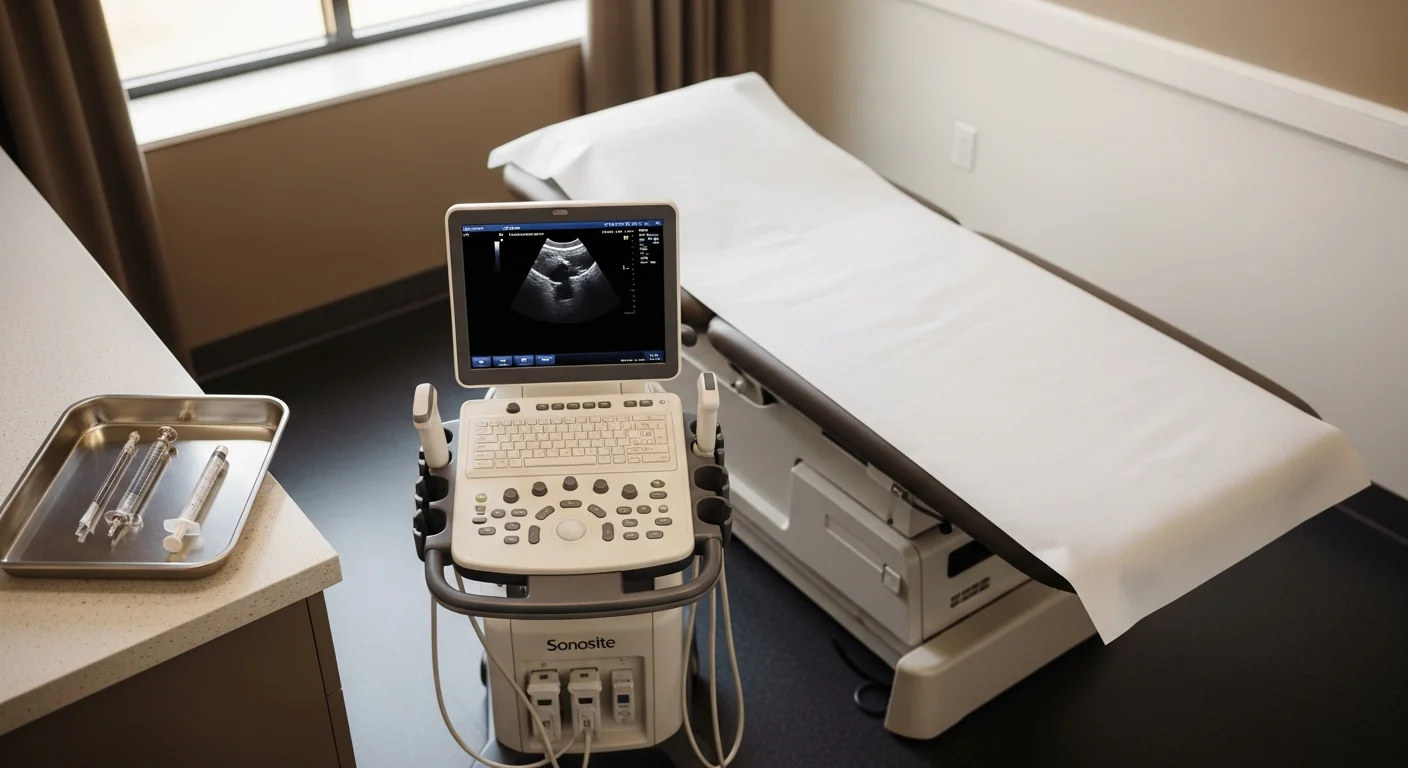
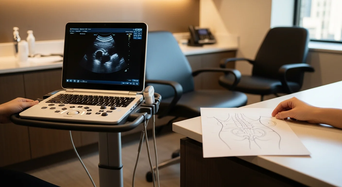
The diagnostic procedure is an ultrasound-guided rectus sheath block at the Carnett’s-positive level. Performed in our procedure suite at 369 Lexington Avenue. Total visit time including the Carnett’s test: about 30 minutes.
Technique: with the patient supine, the abdominal wall anatomy is identified by ultrasound — external oblique, internal oblique, transversus abdominis, and the lateral edge of the rectus sheath at the painful level. A 22-gauge needle is advanced under direct ultrasound visualization to the plane superficial to the posterior rectus sheath at the lateral border of the rectus, at the Carnett’s-positive level. After negative aspiration, 2–3 mL of 0.5% bupivacaine plus 20–40 mg of triamcinolone is delivered, with spread observed in real time.
The diagnostic effect arrives within 5–10 minutes. ≥50% pain abolition is a positive block; ≥75% is highly specific. The patient keeps a pain diary every 30 minutes for the next four hours and at intervals over the next several days.
The therapeutic effect with corticosteroid added typically lasts 6–12 weeks. Patients with clear positive response and diminishing duration are candidates for pulsed radiofrequency ablation of the affected branch — 6–12 months of relief per treatment.
Treatment ladder and when surgery is the right call
Step 1 — neuropathic-pain medication trial. Gabapentin or pregabalin at standard titration. Topical 5% lidocaine patch over the Carnett’s-positive territory is highly effective for focal cutaneous pain.
Step 2 — ultrasound-guided diagnostic and therapeutic rectus sheath block. The workhorse. Diagnostic in 5–10 minutes, therapeutic for 6–12 weeks. Repeatable.
Step 3 — ultrasound-guided hydrodissection. For nerves entrapped in dense fascial scar — particularly common after multiple prior abdominal surgeries. Volume injection of saline or 5% dextrose mechanically separates the nerve from the fibrotic plane.
Step 4 — pulsed radiofrequency ablation. For chronic refractory cases with documented positive blocks and diminishing therapeutic duration. Pulsed (not conventional thermal) — the territory is cosmetically sensitive and the lesion must be neuromodulatory rather than destructive.
Step 5 — peripheral nerve stimulation referral. Percutaneous PNS at the affected branch for severe refractory disease.
Step 6 — anterior neurectomy by peripheral nerve subspecialty. Surgical transection of the anterior cutaneous branch at the rectus sheath, with implantation of the proximal stump into muscle to prevent recurrent neuroma. The Boelens and van Assen Dutch ACNES program publications report 70–85% meaningful pain relief at 12-month follow-up. Performed by peripheral nerve subspecialists within general surgery or plastic surgery. Modal Pain coordinates the referral directly and provides the documented diagnostic-block response surgical programs require.
The ladder is heavily front-loaded toward conservative care because most ACNES responds to repeat therapeutic blocks. Surgery is reserved for the refractory minority.
For referring physicians (especially GI)
When you have a chronic abdominal pain patient with negative endoscopy, negative colonoscopy, negative cross-sectional imaging, and a focal point reproducing the pain on physical exam, run a Carnett's test in your office — 30 seconds, no equipment. A positive test makes ACNES the leading hypothesis. Send the patient for an ultrasound-guided rectus sheath block. Send us: the prior GI workup summary, any prior surgical history (especially laparoscopic port placement records), the medication trial history, and a note on the cutaneous distribution of the pain. We document the Carnett's response at the consultation, run the diagnostic block in the room, and send notes back to your office after each visit. For refractory cases, we coordinate the anterior neurectomy referral.
Ready to evaluate your chronic abdominal pain
If your abdominal pain has persisted through multiple negative GI workups and you can press a specific point to reproduce it, the next step is a Carnett’s test and an ultrasound-guided rectus sheath block. Same-week new-patient consultations are available at 369 Lexington Avenue in Midtown Manhattan.
Verify your insurance covers an ACNES workup Book a same-week diagnostic block
Or call (646) 290-6660.
For the broader framework on peripheral nerve entrapments, see the peripheral nerve entrapment page.
Frequently Asked Questions
ACNES stands for anterior cutaneous nerve entrapment syndrome. The anterior cutaneous branches of the lower thoracic intercostal nerves (T7–T12) pierce the lateral edge of the rectus sheath and turn 90° to supply the overlying abdominal skin. At that 90° turn, the nerve is mechanically vulnerable — by fascial scar from prior surgery, by neuroma after laparoscopic port placement, by hernia of fat through the nerve foramen, or idiopathically. The result is focal anterior abdominal wall pain that mimics intra-abdominal pathology but originates from the abdominal wall itself.
A bedside maneuver that takes 30 seconds and separates abdominal wall pain from intra-abdominal causes. With the patient supine, press firmly on the most painful spot until they confirm reproduction of their typical pain. Then have them lift their head and shoulders off the table — tensing the rectus abdominis — while you maintain the same pressure. If the pain worsens (tensing the wall amplifies it), the source is the abdominal wall — positive Carnett's, ACNES until disproved. If the pain eases or stays the same, the source is intra-abdominal — negative Carnett's, the workup stays with GI/gynecology. The test is documented in the original Boelens 2011 Annals of Surgery paper and in Carnett's original 1926 description.
Three reasons. First, the differential default for chronic abdominal pain is gastrointestinal — endoscopy, colonoscopy, CT abdomen, gastroenterology consult. A negative workup yields a diagnosis of 'IBS' or 'functional abdominal pain.' Neither addresses an abdominal-wall nerve generator. Second, the right diagnostic test (Carnett's plus an ultrasound-guided rectus sheath block) is performed in pain medicine practices, not in GI clinics — the patient who needs the test is rarely referred for it. Third, ACNES mimics conditions that are far more commonly diagnosed (IBS, endometriosis, chronic pelvic pain). Bayesian reasoning makes those the leading hypothesis even when the actual prevalence of ACNES in chronic-abdominal-pain populations is 10–30% per the Dutch ACNES research program (Boelens, van Assen).
An ultrasound-guided rectus sheath block at the lateral edge of the rectus sheath at the Carnett's-positive level. 2–3 mL of 0.5% bupivacaine plus 20–40 mg of triamcinolone deposited superficial to the posterior rectus sheath, where the anterior cutaneous branch makes its 90° turn. The diagnostic effect is immediate — symptoms abolish within 5–10 minutes in a positive case. With corticosteroid added, the therapeutic effect typically lasts 6–12 weeks. Most patients respond to repeat therapeutic blocks; refractory cases are candidates for hydrodissection, pulsed RFA, or surgical neurectomy.
Anterior neurectomy is surgical transection of the anterior cutaneous branch at the rectus sheath, performed for refractory ACNES after the conservative ladder has been exhausted. The published case series from the Dutch ACNES research program (Boelens 2011, van Assen 2015 JAMA Surgery) report 70–85% meaningful pain relief at 12-month follow-up. The procedure is typically performed by a peripheral nerve subspecialist within general surgery or plastic surgery. Modal Pain coordinates the referral and provides the diagnostic-block documentation surgical programs require.
Yes, and this is one reason the diagnosis gets delayed. Patients can have both an intra-abdominal source of pain (IBS, endometriosis, adhesions) and a coexisting abdominal-wall nerve generator. The Carnett's test and diagnostic block isolate the ACNES component. Treating it can produce meaningful relief even when the intra-abdominal condition remains active — the two pain sources are mechanistically independent.
Most commercial PPO plans cover ultrasound-guided rectus sheath blocks for ACNES, typically with prior authorization. Modal Pain verifies your benefits before the first visit. We accept most major commercial PPO plans and do not participate with Medicare or Medicaid. <a href="/verify-insurance/">Check your plan</a> or call (646) 290-6660.