If you have burning or electric pain on the back of your hand and the side of your thumb that started after a wrist injury, after a distal radius surgery, after a de Quervain’s release, after wearing a tight watch or fitness tracker, or — sometimes — for no clear reason at all, and you have been told it is carpal tunnel even though the splinting and the steroid injection at the wrist haven’t helped, you are probably reading the right post. The condition is Wartenberg’s syndrome. A 15-minute ultrasound-guided block confirms it and treats it on the same visit.
Why this gets missed
The clinical territory of Wartenberg’s syndrome overlaps superficially with carpal tunnel — both involve hand pain in a working-age patient — so the differential default is carpal tunnel. Most internists, family physicians, and even many hand specialists default to electrodiagnostic studies and night splinting before performing a careful cutaneous map. The night splint doesn’t help (because the median nerve isn’t the problem), the EMG looks normal or shows minimal median slowing (which is incidental), and the patient ends up either getting a carpal tunnel release that doesn’t help or being told the diagnosis is unclear.
Lanzetta and Foucher’s 1993 case series in International Orthopaedics documented 52 cases — they laid out the diagnostic anchor and the surgical decompression approach. Dellon and Mackinnon’s 1986 Journal of Hand Surgery paper expanded the anatomic and surgical descriptions. Sandvall et al. published a modern 2020 review in the same journal. The condition is real, well-described, and reproducible — and dramatically under-recognized.
The fix is the cutaneous map. Where exactly is the pain? If the patient draws their pain on the back of the hand, the dorsoradial wrist, and the dorsum of the thumb — and explicitly excludes the palm — carpal tunnel is the wrong diagnosis. Wartenberg’s becomes the leading hypothesis. The Tinel test at the brachioradialis tendon 9 cm proximal to the radial styloid is the confirming bedside maneuver. The ultrasound-guided block is the definitive test.
The anatomy that matters
The radial nerve divides into the deep branch (posterior interosseous nerve, motor) and the superficial radial nerve (SRN) in the proximal forearm. The SRN runs along the radial aspect of the forearm, deep to the brachioradialis muscle, accompanied by the radial artery.
Approximately 9 cm proximal to the radial styloid, the SRN emerges from beneath the brachioradialis tendon and pierces the deep fascia to become subcutaneous. From there it crosses over the extensor tendons of the wrist and divides into terminal branches that supply:
- The dorsoradial wrist skin
- The dorsum of the thumb (including the first dorsal web space)
- A patch of skin on the dorsoradial hand
At the emergence point — the brachioradialis-ECRL interval — the nerve is mechanically vulnerable. Three mechanisms produce Wartenberg’s:
- Direct compression from anything sitting on the dorsoradial distal forearm: tight watchband, fitness tracker, handcuffs (occupational pattern in law enforcement), tight athletic wraps.
- Surgical injury during distal radius fixation, de Quervain’s first dorsal compartment release, or wrist arthroscopy where the nerve is retracted, thermally injured by electrocautery, or transected.
- Iatrogenic from IV infiltration — particularly common with antecubital or dorsoradial IV lines that extravasate and produce hematoma compression of the SRN downstream.
The Tinel sign at the brachioradialis-ECRL interval is the single most useful bedside test. Tap at the emergence point and the patient reports a sharp, radiating, electric sensation into the dorsoradial hand and thumb — their typical pain.
How we diagnose
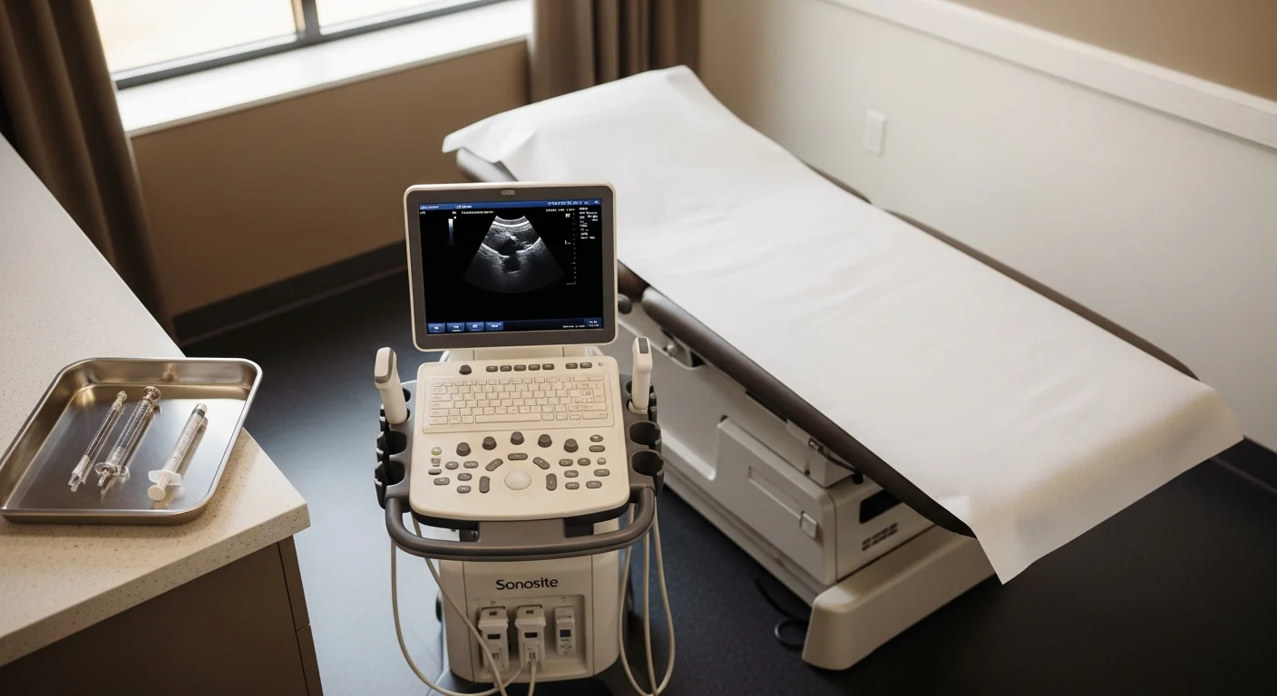
The diagnostic procedure is an ultrasound-guided superficial radial nerve block at the Tinel-positive point. Performed in our procedure suite. Total visit time: about 30 minutes.
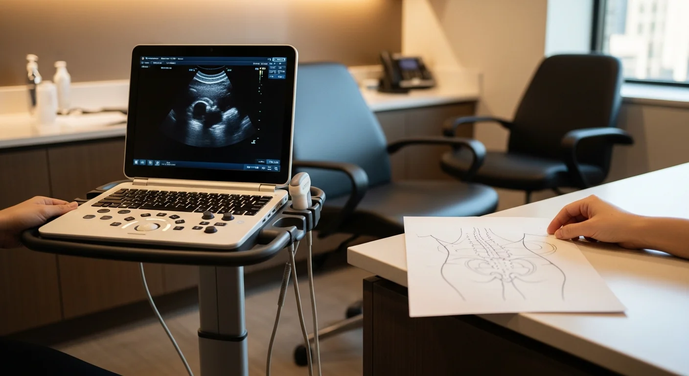
Technique: with the patient’s forearm pronated and supported, the forearm anatomy is identified by ultrasound — brachioradialis muscle and its tendon, extensor carpi radialis longus (ECRL), the radial artery as a landmark, and the SRN as a small hypoechoic structure between the brachioradialis tendon and the ECRL approximately 9 cm proximal to the radial styloid. A 25-gauge needle is advanced under direct ultrasound visualization to the interval between the two muscles, immediately adjacent to the nerve. After negative aspiration, 2–3 mL of 0.5% bupivacaine plus 20 mg of triamcinolone is delivered, with spread observed in real time.
The diagnostic effect arrives within 5–10 minutes. ≥75% relief of the typical dorsoradial burning is the definitive test for Wartenberg’s syndrome. The therapeutic effect with corticosteroid added typically lasts 6–12 weeks. Patients keep a pain diary every 30 minutes for four hours.
Treatment ladder and when surgery is the right call
Step 1 — remove the mechanical compressor. Tight watches, fitness trackers, handcuffs, tight athletic wraps — anything sitting over the brachioradialis-ECRL interval. Cases driven primarily by recent compression often resolve completely at Step 1.
Step 2 — neuropathic-pain medication trial. Gabapentin or pregabalin at standard titration. Topical 5% lidocaine patch over the affected territory.
Step 3 — ultrasound-guided diagnostic and therapeutic SRN block. The workhorse. Diagnostic in 5–10 minutes, therapeutic for 6–12 weeks.
Step 4 — ultrasound-guided hydrodissection. For nerves entrapped in dense post-surgical scar (post-de-Quervain’s release, post-distal-radius fixation). Volume injection of saline or 5% dextrose mechanically separates the nerve from the fascial plane.
Step 5 — pulsed radiofrequency ablation of the SRN. For chronic refractory cases with documented positive blocks. Pulsed (not thermal) — the territory is cosmetically sensitive and the lesion must be neuromodulatory.
Step 6 — surgical decompression or neuroma management. Refractory cases get referred for one of two procedures, depending on whether a discrete neuroma is present:
- Dellon-style external neurolysis of the SRN at the brachioradialis-ECRL interval, performed by hand surgery or peripheral nerve plastic surgery. For patients with chronic mechanical compression without imaging-confirmed neuroma.
- Neuroma excision with RPNI or TMR, performed by peripheral nerve plastic surgery. For patients with established neuroma (often post-surgical or post-IV-infiltration). The proximal nerve stump is implanted into a small denervated muscle graft (RPNI) or coapted to a motor nerve of a small expendable muscle (TMR) to prevent recurrent neuroma formation. Published outcomes for RPNI/TMR in upper-extremity neuroma management are strong.
Modal Pain coordinates these referrals directly and provides the surgical team with the documented diagnostic-block response.
For referring physicians (especially hand surgery)
When you have a patient with persistent dorsoradial hand and thumb pain after distal radius fixation, de Quervain's release, or wrist arthroscopy — and the workup for the original indication looks clean — send: the operative report from the wrist procedure, the prior medication trial history, and a note on the cutaneous distribution of the pain (especially whether it crosses into the palm — if no, Wartenberg's is the leading hypothesis). We document the Tinel sign at the brachioradialis-ECRL interval, run an ultrasound-guided SRN block in the room, and send notes back to your office after each visit. For refractory cases with documented neuroma, we coordinate the RPNI or TMR referral.
Ready to evaluate your dorsoradial wrist and thumb pain
If you have burning or electric pain on the back of the hand and side of the thumb — and a wrist splint or carpal tunnel workup hasn’t helped — the next step is an ultrasound-guided SRN block. Same-week new-patient consultations are available.
Verify your insurance covers a Wartenberg’s syndrome workup Book a same-week diagnostic block
Or call (646) 290-6660.
For the broader framework on peripheral nerve entrapments, see the peripheral nerve entrapment page.
Frequently Asked Questions
No — and confusing the two is the most common reason Wartenberg's gets misdiagnosed for years. Carpal tunnel is median nerve compression at the wrist, producing pain and numbness in the palm of the thumb, index, middle, and radial half of the ring finger. Wartenberg's is superficial radial nerve compression in the distal forearm, producing pain on the back of the hand, the dorsoradial wrist, and the dorsum of the thumb. If your pain is on the back of the hand and NOT in the palm, carpal tunnel is the wrong diagnosis. The territories don't overlap.
Mechanical compression of the superficial radial nerve as it emerges from beneath the brachioradialis tendon about 9 cm proximal to the radial styloid. The nerve becomes subcutaneous at that point and is vulnerable to anything that compresses the dorsoradial forearm: a tight watchband, a fitness tracker, handcuffs (a published occupational pattern in law enforcement), distal radius fracture fixation, de Quervain's release surgery where the nerve is retracted or thermally injured, IV infiltration that produces hematoma compression, repetitive pronation-supination at work, or idiopathic entrapment in patients without any clear trigger.
Ultrasound-guided injection at the brachioradialis-ECRL interval approximately 9 cm proximal to the radial styloid — the Tinel-positive point. 2–3 mL of 0.5% bupivacaine plus 20 mg of triamcinolone deposited around the superficial radial nerve immediately deep to the brachioradialis tendon. The diagnostic effect arrives within 5–10 minutes — abolition of the burning dorsoradial pain is the definitive test. With corticosteroid added, the therapeutic effect typically lasts 6–12 weeks.
Surgical decompression is the salvage option for patients who fail medication, image-guided block + steroid, hydrodissection, and pulsed RFA. The procedure is a Dellon-style external neurolysis of the superficial radial nerve at the brachioradialis-ECRL interval, performed by hand surgery or peripheral nerve plastic surgery. For patients with established neuroma after distal radius surgery or de Quervain's release, neuroma excision with RPNI (regenerative peripheral nerve interface) or TMR (targeted muscle reinnervation) is the modern surgical answer — both techniques implant the proximal nerve stump into a small denervated muscle graft to prevent recurrent neuroma. Modal Pain coordinates the referral and provides the diagnostic-block documentation surgical programs require.
Sometimes, especially if the watch has been the only trigger and the nerve hasn't been chronically irritated for long. Cases driven primarily by recent mechanical compression (a new tight watch, a tight fitness tracker, handcuff exposure) often resolve completely once the compressor is removed. Cases driven by post-surgical neuroma, IV infiltration history, or chronic compression for years rarely resolve from removing the trigger alone — the nerve has been sensitized. The first move is always to identify and remove any active mechanical compressor; the second move is the diagnostic block.
De Quervain's is inflammation of the first dorsal compartment of the wrist — the abductor pollicis longus and extensor pollicis brevis tendons. Pain is localized to the radial side of the wrist over those tendons, and is reproduced by the Finkelstein test (thumb in fist, ulnar deviation). Wartenberg's is nerve pain — burning, electric — and the Tinel sign is positive over the brachioradialis tendon proximal to the wrist. The two can coexist (and post-de-Quervain-release Wartenberg's is a documented iatrogenic complication), but the diagnostic block separates them: an ultrasound-guided SRN block abolishes Wartenberg's pain and does not change de Quervain's pain.
Most commercial PPO plans cover ultrasound-guided superficial radial nerve blocks for Wartenberg's syndrome, typically with prior authorization. Modal Pain verifies your benefits before the first visit. We accept most major commercial PPO plans and do not participate with Medicare or Medicaid. <a href="/verify-insurance/">Check your plan</a> or call (646) 290-6660.