Plantar fasciitis is the most common cause of heel pain in adults, affecting an estimated 2 million Americans each year and roughly 1 in 10 people over a lifetime. It is also one of the most over-diagnosed and under-treated conditions in foot care — many cases of “plantar fasciitis” are actually mimicking conditions (Baxter’s neuropathy, calcaneal stress fracture, fat pad atrophy, tarsal tunnel, S1 radiculopathy) that require completely different treatment.
At Modal Pain Management in Midtown Manhattan, Dr. Alex Movshis uses a focused clinical exam, bedside ultrasound, and an evidence-based, image-guided treatment ladder to diagnose and treat plantar fasciitis correctly the first time. PRP (platelet-rich plasma) is the preferred first-line injection for chronic cases because it addresses the underlying tendinopathy rather than just suppressing inflammation; ultrasound-guided corticosteroid injection is used selectively for acute, severe pain. Most patients are evaluated, diagnosed, and (when appropriate) treated in a single office visit.
This page covers what plantar fasciitis actually is, the conditions it is most commonly confused with, the role of ultrasound in diagnosis, the evidence-based treatment ladder, and why image guidance and PRP-first philosophy produce better long-term outcomes than the historical default of repeated blind cortisone injections.
What Is Plantar Fasciitis?
Plantar fasciitis is a degenerative injury — a tendinopathy — of the plantar fascia, the thick band of connective tissue that runs along the bottom of the foot from the heel bone (calcaneus) to the base of the toes. The injury occurs at or near the medial calcaneal tubercle, where the fascia attaches to the inside-bottom of the heel bone, and is driven by repeated tensile load (walking, running, standing) combined with insufficient recovery and (often) calf tightness or footwear problems.
Despite the historical name “fasciitis” (which implies inflammation), modern histological studies show that established plantar fasciitis is primarily a tendinosis — disorganized collagen fibers, microtears, and abnormal blood vessel ingrowth — rather than active inflammation. This is why pure anti-inflammatory treatment (NSAIDs, repeated cortisone) often provides incomplete or short-lived relief, and why treatments that stimulate tendon healing (PRP, eccentric loading, shockwave therapy) are increasingly favored in the modern evidence base.
The classic patient is a runner, dancer, retail or restaurant worker, parent on their feet all day, or someone with a recent change in activity, footwear, or bodyweight. Peak incidence is age 40–60. The hallmark symptom is sharp medial heel pain with the first steps in the morning or after prolonged sitting, which improves after a few minutes of walking, then often returns after prolonged standing or activity later in the day.
Plantar Fasciitis vs. Other Causes of Heel Pain
Heel pain has several possible causes that look similar on the surface but require completely different treatment. At Modal Pain Management we systematically rule out each one:
- Plantar fasciitis — sharp medial heel pain, worst with the first steps in the morning, tenderness directly over the medial calcaneal tubercle.
- Calcaneal stress fracture — diffuse heel pain that worsens with weight-bearing throughout the day (rather than improving after the first few steps), positive squeeze test, often a recent increase in running mileage. MRI confirms.
- Fat pad atrophy — central (rather than medial) heel pain in older patients or after repeated cortisone injections; the fat pad feels thin on exam.
- Baxter’s neuropathy (first branch of the lateral plantar nerve) — chronic medial heel pain that does not respond to plantar fasciitis treatment, positive Tinel’s sign over the nerve as it passes between the abductor hallucis and quadratus plantae. Frequently misdiagnosed as treatment-resistant plantar fasciitis.
- Tarsal tunnel syndrome — burning, tingling, or numbness on the bottom of the foot from compression of the posterior tibial nerve at the medial ankle.
- S1 radiculopathy (sciatica) — heel and lateral foot pain referred from the S1 nerve root, with associated low back pain, calf pain, weakness in plantar flexion, and an absent ankle reflex.
- Retrocalcaneal bursitis or insertional Achilles tendinopathy — pain at the back of the heel rather than the bottom.
- Posterior tibial tendinopathy — pain along the inside of the foot and ankle in patients with progressive arch collapse (acquired flat foot).
A focused exam plus bedside ultrasound resolves most of these on the first visit. MRI is reserved for atypical or refractory cases.
Diagnosis at Modal Pain Management
A first-visit plantar fasciitis evaluation at Modal Pain Management includes:
- Focused history — onset (acute vs. insidious), pain pattern (first-step pain in the morning is the hallmark), recent changes in activity, footwear, or bodyweight, prior treatments and their effect, and any back or calf symptoms that might suggest a different diagnosis.
- Physical examination — inspection of the foot and arch (high-arch vs. flat foot), palpation of the medial calcaneal tubercle (the classic tender point), windlass test (passive dorsiflexion of the great toe reproduces heel pain in plantar fasciitis), Tinel’s sign over the tibial nerve and Baxter’s nerve, calf flexibility (Silfverskiöld test), and a focused neurological exam to rule out S1 radiculopathy.
- Bedside ultrasound — visualization of the plantar fascia at the medial calcaneal tubercle. A normal fascia is 2–4 mm thick; a thickness greater than 4 mm is consistent with plantar fasciitis, and greater than 5 mm typically indicates more severe disease that benefits from injection or shockwave. Ultrasound also identifies hypoechoic areas of tendinosis, partial tears, calcaneal spurs, neovascularization, and — critically — guides the injection if one is performed.
- Targeted imaging — X-ray if there is concern for a calcaneal stress fracture or large bone spur; MRI for atypical presentations, suspected stress fracture, or before considering surgery.
The single-visit diagnostic workflow with bedside ultrasound is one of the main reasons patients with chronic, undifferentiated heel pain often achieve faster definitive answers at Modal Pain Management than through serial referral.
Image-Guided Treatment Ladder
The evidence-based treatment for plantar fasciitis follows a structured ladder. About 80–90% of patients resolve in the first 1–2 steps; only a small minority need to escalate to injection or beyond.
Step 1: Conservative Treatment (Weeks 0–6)
For most patients with acute or mild symptoms, the first 4–6 weeks emphasize structured stretching, footwear correction, and activity modification:
- Plantar fascia-specific stretching (DiGiovanni protocol) — sit with the affected leg crossed over the other, pull the toes back toward the shin until you feel a stretch in the arch, hold for 10 seconds, repeat 10 times, perform 3 times daily (especially before getting out of bed in the morning). This has the strongest single-intervention evidence base for plantar fasciitis.
- Calf stretching — gastrocnemius (knee straight) and soleus (knee bent) stretches, 30 seconds, 3 sets, 2–3 times daily.
- Supportive footwear — replace flat or worn-out shoes with supportive shoes appropriate for the patient’s foot type. Avoid going barefoot on hard surfaces.
- Over-the-counter or prescription orthotics — heel cups, arch supports, or custom orthotics matched to the foot.
- Activity modification — temporary reduction in running mileage, intensity, and standing time; cross-train with low-impact exercise (swimming, cycling, elliptical).
- NSAIDs for short-term symptom control if you tolerate them (naproxen 500 mg twice daily for 7–10 days, with food).
- Night splint — particularly helpful for patients with severe morning heel pain; holds the foot in a stretched position overnight.
About 80–90% of plantar fasciitis cases resolve with this approach within 6–12 weeks.
Step 2: PRP (Platelet-Rich Plasma) Injection — Preferred First Injection (Weeks 6–12+)
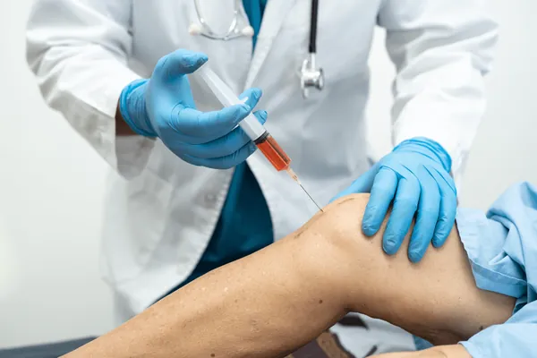
For patients whose pain persists beyond 4–6 weeks of structured conservative care, or for chronic plantar fasciitis on ultrasound (fascia thickness >4 mm with hypoechoic changes), PRP injection is the preferred first injection in the modern evidence base. PRP uses a small sample of the patient’s own blood, processed in-office to concentrate the platelets, then injected directly into the damaged plantar fascia under real-time ultrasound guidance. The growth factors released by the platelets stimulate genuine tendon healing rather than simply suppressing inflammation.
PRP takes longer to work than cortisone (full effect at 6–12 weeks, with a temporary increase in soreness in the first 1–2 weeks as the healing response begins), but multiple randomized trials show 70–85% of patients with chronic plantar fasciitis achieve durable relief at 12+ months — substantially better long-term outcomes than cortisone. PRP is typically not covered by insurance and has an out-of-pocket cost, which we discuss transparently before scheduling.
Step 3: Ultrasound-Guided Corticosteroid Injection (Selected Cases)
Ultrasound-guided corticosteroid injection is appropriate in selected scenarios — typically acute, severe pain where a patient absolutely must function in the next 2–4 weeks (a critical work or athletic event). Cortisone provides faster initial relief (1–2 weeks to peak effect) but is associated with higher 6- and 12-month recurrence rates. Ultrasound guidance is essential: blind heel injections frequently miss the fascia and deposit medication in the fat pad, which both reduces effectiveness and increases the risk of fat pad atrophy and plantar fascia rupture (a small but real risk of about 2–10% in older studies of repeated blind injection). We do not repeat cortisone injections in the plantar fascia within 3–6 months and do not perform more than 2–3 injections per fascia over the patient’s lifetime.
Step 4: Extracorporeal Shockwave Therapy (ESWT) for Refractory Cases
For chronic, refractory plantar fasciitis (>6 months) that has not adequately responded to conservative care and injection, extracorporeal shockwave therapy (ESWT) is an evidence-based next step. Shockwave delivers controlled high-energy acoustic waves to the plantar fascia, stimulating a healing response without the need for injection. ESWT has good evidence in chronic plantar fasciitis, with success rates of 60–80% in patients who have failed first-line injection. It is performed in 2–3 sessions over 2–4 weeks. We coordinate ESWT referral with experienced operators in NYC and integrate it into a comprehensive treatment plan.
When Surgery Is Considered
Surgical plantar fascia release (open or endoscopic) is reserved for the small subset of patients (<5%) with chronic, severe plantar fasciitis that has not responded to 6–12 months of structured non-operative treatment including PRP and shockwave. We coordinate referrals to NYC orthopedic foot-and-ankle specialists when surgery becomes appropriate — but the great majority of patients avoid this pathway.
Physical Therapy and Self-Care
Image-guided injection accelerates pain relief, but durable recovery requires addressing the underlying fascia, calf, and biomechanics. A targeted plantar fasciitis program typically includes:
- Plantar fascia-specific stretching (DiGiovanni protocol) — the single highest-evidence intervention; performed 3 times daily including before getting out of bed in the morning.
- Calf stretching — gastrocnemius and soleus, twice daily.
- Eccentric calf strengthening — heel drops on a step, slow controlled lengthening contractions, 3 sets of 15.
- Intrinsic foot strengthening — toe scrunches with a towel, marble pickups, short-foot exercise.
- Footwear and orthotic correction — supportive shoes with appropriate arch support, prescription orthotics for severe pes planus or pes cavus, avoidance of going barefoot or wearing flip-flops on hard surfaces.
- Training-load management for runners — avoid sudden increases in mileage or intensity, follow the 10% rule (no more than 10% increase in weekly mileage), build in proper recovery.
- Self-care — frozen water bottle rolling under the arch for 10 minutes after activity, ice for 15 minutes after long days on the feet, night splint for severe morning pain.
Most patients see meaningful improvement within 6–12 weeks of starting structured rehab combined with appropriate injection.
When to Seek Specialist Care
See a pain specialist for plantar fasciitis evaluation if any of the following apply:
- Heel pain that has not improved with 4–6 weeks of plantar fascia stretching, calf stretching, supportive footwear, and activity modification.
- Pain that is interfering with work, sleep, or daily activities (taking the first steps in the morning, walking the dog, standing at work).
- Symptoms that have been present for more than 3 months despite home treatment.
- A previous episode that resolved and has now recurred.
- Pain that is atypical (central rather than medial heel pain, pain that worsens through the day rather than improving with the first few steps, associated back or calf symptoms, burning or tingling sensation) — these patterns suggest a different diagnosis that needs to be ruled out.
Red-flag symptoms — sudden severe heel pain after a “pop” while walking or running (suspicious for plantar fascia rupture), inability to bear weight, fever or warmth in the heel (concern for infection or septic joint), or numbness in the foot — require prompt evaluation rather than an outpatient plantar fasciitis workup.
Why Modal Pain Management for Plantar Fasciitis in NYC
Modal Pain Management is a focused, physician-owned interventional pain practice in Midtown Manhattan. Dr. Alex Movshis is dual board-certified in Anesthesiology and Pain Medicine and completed an ACGME-accredited interventional pain medicine fellowship at the Icahn School of Medicine at Mount Sinai. NPI 1942741160 — see our evidence and credentials page and the physician bio for full verification.
Three things differentiate plantar fasciitis care at Modal Pain Management:
- Single-visit diagnosis with bedside ultrasound. A focused exam plus real-time ultrasound imaging of the plantar fascia — at the consultation visit — eliminates the back-and-forth of separate imaging appointments, measures fascia thickness objectively, and screens for the mimickers (Baxter’s neuropathy, calcaneal stress fracture, S1 radiculopathy) that are commonly missed in patients treated for plantar fasciitis without lasting relief.
- Image-guided, PRP-first treatment philosophy. Every injection is performed under real-time ultrasound guidance — blind heel injections are substantially less accurate and carry higher risks. The evidence base for chronic plantar fasciitis now favors PRP over cortisone for long-term outcomes, and we use PRP as the preferred first-line injection for most patients with chronic disease, reserving cortisone for selected acute scenarios.
- Coordinated medical, interventional, and rehabilitation plan. We work directly with NYC physical therapists who specialize in lower-extremity tendinopathy, coordinate ESWT referral when needed, and build out the full footwear, orthotic, and training-load plan that converts injection benefit into durable recovery.
Office: 369 Lexington Avenue, Floor 25, New York, NY 10017. Same-week appointments available. Most major insurance accepted for diagnostic visits and corticosteroid injections — verify your benefits or call (646) 290-6660 and our team will check coverage for you. PRP and ESWT have associated out-of-pocket costs that we discuss transparently before scheduling.