Modal Pain’s approach to PRP
Platelet-rich plasma is one of the most over-marketed and under-explained procedures in musculoskeletal medicine. At Modal Pain Management, we perform PRP selectively, image-guided, and only for conditions where peer-reviewed evidence supports it. We discuss both the favorable and unfavorable studies before you decide, publish our per-injection pricing transparently, and never package PRP as part of a pre-paid “wellness protocol.” This page is written in the same spirit: honest about what PRP can and cannot do for musculoskeletal pain in 2026.
PRP is an autologous biologic. That means the active ingredient is drawn from your own blood on the day of the procedure and returned to your body after a single centrifuge spin. Because it is autologous, PRP is not regulated by the FDA as a drug — it is regulated as a minimally manipulated same-day tissue product under 21 CFR 1271.15(b). It does not contain steroids, stem cells, amniotic tissue, or any foreign biologic additive.
The biology: what PRP actually delivers
Platelets are best known for clotting, but they also carry more than 300 bioactive molecules in their alpha granules. When platelets are concentrated and delivered to damaged tissue, they release a cascade of growth factors over the first 7–10 days, including platelet-derived growth factor (PDGF), transforming growth factor-beta (TGF-β), vascular endothelial growth factor (VEGF), insulin-like growth factor-1 (IGF-1), basic fibroblast growth factor (bFGF), and epidermal growth factor (EGF).
In a healthy acute injury, this cascade happens naturally. In chronic tendinopathy and in mild-to-moderate osteoarthritis, the local healing response has stalled — the tissue is stuck in a degenerative, failed-healing state rather than progressing to repair. Concentrated PRP delivers a supraphysiologic dose of growth factors to the exact site, with the goal of restarting a productive healing cascade: angiogenesis, tenocyte or chondrocyte anabolic activity, matrix deposition, and collagen remodeling.
This biology matters clinically. PRP is slower than cortisone. It often causes a several-day flare as inflammation is actively triggered. Real benefit — when it occurs — shows up at 4–6 weeks and continues to improve through 3–6 months. PRP is not a quick fix.
Conditions we treat with PRP at Modal Pain
Our offered indications are organized by strength of evidence rather than by marketing appeal.
Tendinopathies with strongest evidence. Lateral epicondylitis (tennis elbow), medial epicondylitis (golfer’s elbow), patellar tendinopathy (jumper’s knee), chronic plantar fasciitis, and select rotator cuff tendinopathies. These are pain syndromes where the pathology is chronic tendinosis — not an acute tear and not primarily inflammatory — and where cortisone has limited durability or is contraindicated due to tendon weakening risk. See tennis elbow and plantar fasciitis for condition-specific discussion.
Mild-to-moderate osteoarthritis (Kellgren–Lawrence 1–3). Knee osteoarthritis and hip osteoarthritis are the best-studied joint indications. The evidence is mixed but increasingly favors PRP over hyaluronic acid and saline at 6–12 months in the subset of patients with preserved joint space and no mechanical deformity. We are transparent: the RESTORE trial (Bennell et al., JAMA 2021) found no PRP benefit over saline in knee OA, and that matters. Other trials and meta-analyses favor PRP. We share both sides. See knee osteoarthritis and hip osteoarthritis.
Ligament and soft-tissue injuries. Partial MCL, UCL, and ankle ligament injuries; mild muscle strains; hip labral pathology in the non-surgical candidate.
Conditions where we will tell you PRP is not the right choice. End-stage osteoarthritis with bone-on-bone changes, complete tendon ruptures, active rotator cuff full-thickness tears in the surgical candidate, active local or systemic infection, radicular pain from spine pathology (epidural steroid injection or radiofrequency ablation is evidence-based), and facet-mediated spine pain (see medial branch blocks).
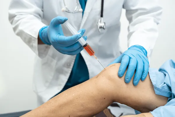
Why image guidance is non-negotiable
Published ultrasound accuracy studies have repeatedly shown that blind landmark injections of the knee, shoulder, hip, and plantar fascia miss the intended target 20–70% of the time depending on operator and joint. For a procedure where the entire mechanism depends on depositing a small volume of concentrated biologic into a specific tendon fiber layer or intra-articular space, blind injection is not an acceptable standard.
Every PRP injection at Modal Pain is performed under real-time ultrasound or fluoroscopic guidance. This allows us to (1) confirm the needle tip is in the pathologic tissue before injection, (2) avoid nerves and vessels, (3) document procedure accuracy in the chart, and (4) reproduce the approach on subsequent injections. If you are considering PRP at a clinic that does not use image guidance, ask why.
The Modal Pain PRP procedure, step by step
Step 1 — Medical clearance and diagnostic review. Before scheduling, we review your imaging (MRI, ultrasound, or X-ray) and rule out contraindications: active infection, thrombocytopenia, anemia, recent cortisone in the same site, or pathology better addressed surgically.
Step 2 — Blood draw (day of procedure). A standard venous draw of 15–60 mL depending on the target. No fasting required. Stay well-hydrated the day before.
Step 3 — Centrifuge and preparation. The sample is spun in a closed, single-use kit in the procedure room. Preparation time is 10–15 minutes. We use single-spin systems that yield a platelet concentration of roughly 3–5× baseline.
Step 4 — Image-guided injection. The skin is prepped with chlorhexidine. The target is identified on ultrasound or fluoroscopy. Local anesthetic is infiltrated at the skin and along the needle track (we intentionally minimize lidocaine at the injection target because high concentrations of lidocaine are cytotoxic to chondrocytes and tenocytes). The PRP is deposited precisely into the pathologic tissue or joint space. The needle is withdrawn.
Step 5 — Post-procedure instructions. You go home the same day. Expect soreness for 3–7 days. No NSAIDs for 2 weeks before and 2 weeks after (NSAIDs can blunt the PRP healing response). Ice only if needed for comfort. Most patients resume desk work the same or next day. Return to loading, sport, or manual labor is diagnosis-specific.
What to expect in the weeks after PRP
- Days 0–7. Soreness, stiffness, mild swelling. This is expected and reflects the intended inflammatory phase of healing. Acetaminophen is allowed. Avoid NSAIDs.
- Weeks 2–4. Pain typically returns to baseline or better. This is not the end-state result.
- Weeks 4–6. First meaningful improvement in most responders.
- Months 3–6. Peak benefit.
- Month 3 reassessment. If there has been no meaningful clinical change at three months, we reassess — repeat injection, switch modality, or re-examine the diagnosis.
How we decide between PRP, corticosteroid, hyaluronic acid, and surgery
A thoughtful diagnosis-specific ladder looks like this:
- Acute or inflammatory joint pain. Cortisone is usually the correct first choice — rapid onset, strong anti-inflammatory effect, covered by insurance. PRP is not an acute anti-inflammatory.
- Chronic tendinopathy not responsive to physical therapy. PRP has strong evidence. Cortisone provides short-term relief but has been shown to weaken tendon structure with repeated use.
- Mild-to-moderate knee OA with symptoms beyond PT. Reasonable options include cortisone (rapid, covered), hyaluronic acid (5 weekly visits, partial insurance coverage), or PRP (self-pay, potentially more durable). We discuss all three.
- End-stage OA with mechanical deformity. Orthopedic referral for joint replacement.
- Spine pain. PRP is not the evidence-based choice. See epidural steroid injections, medial branch blocks, and radiofrequency ablation.
Book an honest PRP consultation
Modal Pain Management is located at 369 Lexington Avenue, Floor 25, New York, NY 10017. Dr. Alexander Movshis performs all PRP procedures personally under ultrasound or fluoroscopic guidance. Consultation includes a full review of your imaging, an honest discussion of whether PRP is the right choice (it often is not), and — if PRP is appropriate — transparent self-pay pricing with no packages or pressure.
Verify insurance and schedule a consultation or call (646) 290-6660.
Our practice difference
We do not market PRP as a cure-all. We do not bundle it into membership programs. We do not upsell stem cell, amniotic, or unregulated biologic products. We do perform every injection with image guidance, document procedure accuracy, and follow up with a reassessment at three months to decide whether the biology actually worked for you. If a cortisone injection, hyaluronic acid series, or referral to orthopedic surgery is the better answer, we will tell you so — even if it means you pay us less.
Selected references
Mishra AK et al. Efficacy of platelet-rich plasma for chronic tennis elbow: a double-blind, prospective, multicenter, randomized controlled trial. Am J Sports Med. 2014;42(2):463-471.
Patel S et al. Treatment with platelet-rich plasma is more effective than placebo for knee osteoarthritis: a prospective, double-blind, randomized trial. Am J Sports Med. 2013;41(2):356-364.
Krogh TP et al. Treatment of lateral epicondylitis with platelet-rich plasma, glucocorticoid, or saline: a randomized, double-blind, placebo-controlled trial. Am J Sports Med. 2013;41(3):625-635.
Bennell KL et al. Effect of intra-articular platelet-rich plasma vs placebo injection on pain and medial tibial cartilage volume in patients with knee osteoarthritis: the RESTORE randomized clinical trial. JAMA. 2021;326(20):2021-2030.
Filardo G et al. Platelet-rich plasma intra-articular knee injections show no superiority versus viscosupplementation: a randomized controlled trial. Am J Sports Med. 2015;43(7):1575-1582.
Andia I, Maffulli N. Platelet-rich plasma for managing pain and inflammation in osteoarthritis. Nat Rev Rheumatol. 2013;9(12):721-730.