Knee osteoarthritis is the single most common cause of disabling adult knee pain and one of the leading causes of mobility loss worldwide. At Modal Pain Management in Midtown Manhattan, Dr. Alex Movshis uses high-resolution imaging, a structured physical examination, and image-guided diagnostic and therapeutic injection to deliver a modern, knee-preservation approach to knee arthritis — treating the disease early, slowing its progression, and reserving surgery for the patients who actually need it. Most patients walk out of the consultation visit with a clear diagnosis, a written treatment plan, and a meaningfully better answer than “you’ll need a knee replacement someday.”
This page covers what knee osteoarthritis actually is, how it is reliably distinguished from the dozen other things that can cause knee pain, how Dr. Movshis diagnoses it, the evidence-based image-guided non-surgical treatment ladder we use — including the most powerful interventional option for moderate-to-advanced disease, image-guided genicular nerve radiofrequency ablation — when surgical referral is appropriate, and what to expect at your first visit.
Knee Osteoarthritis Is Not an Automatic Walk to the Operating Room
The dominant clinical narrative around knee osteoarthritis for the past three decades has been: conservative treatment fails, you get total knee replacement (TKA), end of story. Modern knee-preservation pain management has changed that calculus. There is now a structured, evidence-based, image-guided ladder of non-surgical treatments — corticosteroid injection, hyaluronic acid viscosupplementation (the “gel” injection), platelet-rich plasma (PRP), genicular nerve block and radiofrequency ablation, weight management, and targeted physical therapy — that, applied early and in the right sequence, can extend the useful life of the native knee by years for the right patients.
This matters for two reasons. First, knee implants have a finite lifespan (typically 15–25 years depending on age, activity level, and implant type), and revision arthroplasty is technically harder and biomechanically less successful than the index procedure — so delaying replacement is intrinsically valuable for younger and middle-aged patients, particularly those in their 50s and early 60s who could otherwise outlive their first implant. Second, many patients told they need knee replacement on the basis of an X-ray finding alone are actually candidates for several more years of high-quality function on a structured non-surgical program. Dr. Movshis works alongside NYC-based orthopedic surgeons and refers when surgical replacement is the right answer — and helps patients delay it when it is not.
A third population that frequently benefits from the structured non-surgical ladder: patients with significant medical comorbidities (cardiac disease, diabetes, anticoagulation, advanced age) who carry high surgical risk and for whom image-guided intra-articular injection plus genicular nerve RFA can deliver knee-replacement-level pain control without an operating room.
Why It Hurts There: The Anatomy of Knee Osteoarthritis
The knee is a complex hinge-and-pivot synovial joint with three articulating compartments: the medial tibiofemoral compartment (the inside of the knee where the femur meets the tibia), the lateral tibiofemoral compartment (the outside), and the patellofemoral compartment (where the kneecap glides on the femur). All articulating surfaces are covered with hyaline cartilage that distributes load and allows nearly frictionless motion. The medial and lateral menisci — C-shaped fibrocartilage shock absorbers — sit between the femur and tibia and absorb 50–70% of the load transmitted across the joint. The synovial membrane lines the joint and produces the synovial fluid that nourishes cartilage. The infrapatellar fat pad (Hoffa’s fat pad) cushions the front of the joint and is densely innervated. Pain from the knee joint is carried by the genicular nerves — sensory branches that arise from the femoral, common peroneal, and tibial nerves and converge on the joint capsule.
In knee osteoarthritis, the hyaline cartilage progressively thins, fissures, and ultimately wears away, exposing the underlying subchondral bone. The medial compartment is involved first in 70–80% of cases (because most people walk with a slight varus moment that loads the medial side preferentially), the patellofemoral compartment second, and the lateral compartment last. As cartilage is lost, the joint space (the gap between femur and tibia on X-ray) narrows. Subchondral bone responds by becoming sclerotic, by forming bony osteophytes (spurs) at the joint margins, and by developing subchondral cysts. The synovium becomes inflamed and produces pain-mediating cytokines, often producing visible effusions. The menisci degenerate and tear (degenerative meniscal tearing is a feature of knee OA, not an independent disease — and is the reason why arthroscopic meniscectomy fails to relieve pain in OA knees, per the landmark Sihvonen and Katz randomized trials). The capsule thickens and the joint loses range of motion. Pain is generated by inflamed synovium, exposed subchondral bone, irritated capsule, and the genicular nerves that innervate the capsule.
Understanding this anatomy explains two clinical realities. First, image-guided intra-articular injection is more accurate than blind landmark injection — particularly when the joint space is narrowed, when there is a small effusion, or when the patient is obese. Second, the genicular nerves carry the pain signal from a degenerated joint to the brain — and disrupting them with radiofrequency ablation interrupts the pain pathway without altering the joint itself. Both image-guided intra-articular treatment and genicular nerve RFA are core to the modern knee-preservation toolkit.
Knee Osteoarthritis vs. The Other Causes of Knee Pain
The single most important step in evaluating knee pain is determining where the pain actually is and what is generating it. The differential diagnosis is wide and the wrong diagnosis leads to the wrong treatment.
The most common cause of generalized knee pain in adults over 50 is knee osteoarthritis itself — covered on this page. But several other conditions cause knee pain, mimic knee OA, or coexist with it:
Knee bursitis is inflammation of one of the small fluid-filled sacs around the knee — most commonly the prepatellar bursa (in front of the kneecap, classically a “carpet layer’s knee” or “housemaid’s knee”), the pes anserine bursa (medial side, just below the joint line), or the infrapatellar bursa (below the kneecap). Bursitis pain is superficial, localized to the bursa, and reproducible with direct palpation — distinct from the deep joint-line pain of knee OA. Importantly, knee bursitis frequently coexists with knee OA in the same patient (particularly pes anserine bursitis on the medial side) and the bursa can be a major contributor to a patient’s symptom burden even when the joint itself is the dominant pathology. Image-guided diagnosis sorts these out and image-guided injection treats them selectively.
Patellofemoral pain syndrome (“runner’s knee” or “anterior knee pain”) is pain at the front of the knee around or behind the kneecap, typically worse with stair descent, prolonged sitting (“theater sign”), and squatting — and most common in younger active patients. Patellofemoral OA is a specific compartment of knee OA that affects the cartilage behind the kneecap and is more common in middle-aged and older patients with prior patellar trauma or maltracking.
Meniscal tear can be acute and traumatic (in younger patients, with a clear injury and true mechanical locking, catching, or giving way) or degenerative (in middle-aged and older patients with no clear injury, where the tear is a feature of underlying knee OA rather than an independent problem). Symptomatic acute traumatic tears in young patients can benefit from arthroscopic repair; degenerative meniscal tears in OA knees do not benefit from arthroscopic meniscectomy in well-conducted RCTs (Sihvonen 2013, Katz 2013) — those patients should be treated with the knee OA ladder.
Iliotibial band (IT band) syndrome causes pain on the lateral side of the knee just above the joint line, classically in runners and cyclists, and is reproducibly tender at the lateral femoral epicondyle. It is not joint pain.
Medial collateral ligament (MCL) and lateral collateral ligament (LCL) injury cause pain along the inside or outside of the knee respectively, typically with a clear injury history and tenderness directly over the ligament.
Pes anserine tendinopathy/bursitis causes medial knee pain just below the joint line and frequently coexists with knee OA — particularly common in overweight middle-aged women.
Baker’s cyst is a fluid-filled outpouching of the posterior knee joint capsule into the popliteal fossa, frequently seen in knee OA. A ruptured Baker’s cyst can mimic deep vein thrombosis (a “pseudo-thrombophlebitis” syndrome) and warrants urgent evaluation.
Crystal arthropathy — gout (monosodium urate) and pseudogout (calcium pyrophosphate) — causes acute, severely inflamed monoarticular knee pain with redness, warmth, and a tense effusion. Diagnosis requires joint aspiration and crystal analysis under polarized microscopy.
Septic arthritis is a surgical emergency. Acute monoarticular knee pain with fever, severe redness, warmth, inability to bear weight, and a tense effusion warrants urgent ED evaluation with joint aspiration, gram stain, and culture.
Inflammatory arthritis — rheumatoid arthritis, psoriatic arthritis, ankylosing spondylitis, IBD-associated arthritis — typically presents with multi-joint involvement, prolonged morning stiffness (more than 60 minutes), elevated inflammatory markers, and characteristic systemic features. Modal Pain coordinates rheumatology co-management when the pattern raises concern.
Referred pain from the hip is a frequently missed cause of “knee pain” — hip osteoarthritis can refer pain into the front of the thigh and occasionally to the knee itself. Patients with knee pain in the absence of clear knee findings should have the hip examined carefully.
Lumbar radiculopathy at L3 or L4 can refer pain to the front of the thigh and the knee. The neurologic exam, the distribution of the pain, and the pattern of mechanical aggravation help distinguish.
Sorting out which of these is generating your knee pain — or which combination, since they frequently coexist — is the diagnostic work of the consultation visit.
How Knee Osteoarthritis Is Diagnosed
The diagnosis of knee osteoarthritis is fundamentally clinical and radiographic — meaning it is made by combining a careful history, a structured physical examination, and a weight-bearing knee X-ray. MRI is rarely needed to make the diagnosis itself; its role is to evaluate specific differential considerations (meniscal pathology in younger patients, suspected osteonecrosis, suspected occult fracture).
The history establishes the pattern: age over 50, slowly progressive activity-related knee pain (typically medial-sided), morning stiffness lasting less than 60 minutes that loosens with movement, mechanical pain worse with weight-bearing and stair use, and frequent prior diagnoses of “wear and tear.” Any history of true mechanical locking, true giving way, prior surgery, prior trauma, prior infection, or systemic inflammatory features is recorded.
The physical examination looks for visible varus or valgus alignment, the presence and size of an effusion (the bulge sign and the patellar tap), joint-line tenderness (medial more than lateral in classic knee OA), patellofemoral crepitus and tenderness, range of motion (early loss of full extension is characteristic), quadriceps strength and bulk, gait pattern, and the response to McMurray testing for meniscal pathology. The contralateral knee, the hip, and the lumbar spine are examined to identify referred and coexisting sources.
A weight-bearing knee X-ray (standing AP, lateral, and skyline patellofemoral views) is the imaging study that confirms the diagnosis. The Kellgren-Lawrence grading scale (grade 0 = no OA, grade 1 = doubtful, grade 2 = mild definite OA, grade 3 = moderate, grade 4 = severe with bone-on-bone changes) is the standard. Joint space narrowing, osteophytes, subchondral sclerosis, and subchondral cysts are the four hallmark findings. Importantly, X-ray findings frequently underestimate symptom severity — patients with grade 2 OA can have substantial pain, and patients with grade 4 OA can sometimes have surprisingly little — so the X-ray informs but does not dictate the treatment plan.
MRI is reserved for specific clinical questions: suspected acute meniscal tear in a younger patient with a true injury, suspected osteonecrosis (subchondral insufficiency fracture of the knee — more common in older women), suspected occult fracture, evaluation of a Baker’s cyst, or pre-operative planning. Routine MRI of every painful knee in an older adult is low-yield and often shows incidental findings (degenerative meniscal tearing, bone marrow edema, small effusions) that do not change management. Modal Pain orders MRI selectively, only when the answer will change what we recommend.
Bedside ultrasound at the consultation visit is used to evaluate the joint for an effusion, to assess the suprapatellar bursa, to screen for a Baker’s cyst, to grade synovitis, and to plan the trajectory of an intra-articular injection — all in real time during the same visit.
A diagnostic image-guided intra-articular knee injection with local anesthetic is the gold-standard test when the source of pain is unclear after history, exam, and X-ray. If at least 75% of the patient’s typical knee pain is reproducibly relieved within the anesthetic window, the knee joint itself is confirmed as the pain generator. This is particularly useful when knee OA coexists with hip pathology, lumbar radiculopathy, or pes anserine bursitis and the relative contribution of each needs to be sorted out before treatment.
Joint aspiration with synovial fluid analysis is added when the picture raises concern for septic arthritis (fever, severe inflammation, inability to bear weight) or crystal arthropathy (acute severely inflamed monoarticular swelling).
Laboratory testing for inflammatory arthritis is added when the pattern is atypical — bilateral symmetric polyarticular involvement, prolonged morning stiffness, elevated inflammatory markers, or characteristic systemic features.
The Image-Guided Non-Surgical Treatment Ladder
Modal Pain Management uses a structured, evidence-based, image-guided knee-preservation ladder. The right step on the ladder depends on disease stage, functional impairment, prior response to treatment, surgical candidacy, and patient goals. The ladder is sequential — each step builds on the foundation of the prior steps.
Step 1 — Foundation: Weight Management Plus Structured Physical Therapy
Every patient with knee osteoarthritis benefits from this foundation, regardless of where they ultimately end up on the ladder. Weight management is the single most powerful disease-modifying intervention in knee OA — a 5–10% reduction in body weight reliably reduces knee pain, improves function, and slows radiographic progression in multiple high-quality randomized trials (the Look AHEAD trial, the Messier IDEA trial). Every pound of body weight produces approximately 3–6 pounds of force across the knee during walking and 6–10 pounds during stair climbing, so even modest weight loss meaningfully unloads the joint. For patients in whom dietary and lifestyle approaches plateau, Modal Pain coordinates medical weight loss — including GLP-1 agonist therapy where indicated — as part of a comprehensive knee-preservation plan.
Structured physical therapy is the single best evidence-based exercise intervention for knee OA — quadriceps and hip abductor strengthening, hamstring and calf flexibility, low-impact aerobic conditioning (stationary cycling, elliptical, aquatic therapy), and gait retraining. The OARSI (Osteoarthritis Research Society International) guidelines and the AAOS clinical practice guidelines both rank PT and weight management as Tier 1 interventions. Many patients improve substantially with this foundation alone — and even patients who progress further on the ladder benefit because injection-driven pain control enables more productive PT.
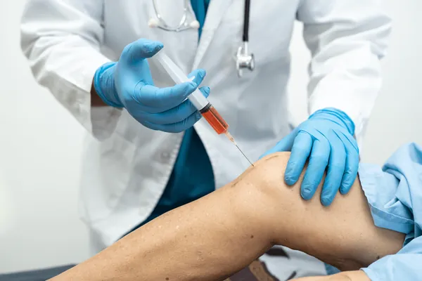
Step 2 — Image-Guided Intra-Articular Corticosteroid Injection
For acute flares, for fast functional improvement before a major life event, and as a diagnostic tool to confirm the knee joint as the pain generator, image-guided intra-articular corticosteroid injection is the workhorse of the early-to-mid ladder. Onset of relief is 3–7 days, magnitude of relief is meaningful in 70–80% of patients, and duration is 6–12 weeks on average. The injection is performed under fluoroscopic or ultrasound guidance — image guidance pushes accuracy from 70–85% (blind landmark) to >95%, particularly important when the joint space is narrowed, when there is a small effusion, or in obese patients. Modal Pain limits intra-articular corticosteroid injections to a sustainable cadence (typically no more than 3–4 per year per joint) to avoid the chondrotoxic concerns raised by repeated high-dose steroid exposure (McAlindon JAMA 2017). For patients who need more frequent or more durable control, the ladder progresses to viscosupplementation, PRP, or genicular nerve RFA.
Step 3 — Image-Guided Hyaluronic Acid (Viscosupplementation, “Gel Injection”)
Hyaluronic acid is FDA-approved specifically for knee osteoarthritis and has decades of clinical evidence. The intra-articular injection of high-molecular-weight hyaluronic acid restores the viscoelastic properties of synovial fluid, lubricates the joint, and may have anti-inflammatory effects on the synovium. Multiple FDA-approved formulations are available — Synvisc, Hyalgan, Euflexxa, Supartz, Orthovisc, Monovisc, Gel-One, GenVisc — delivered as a single injection or a series of 3–5 weekly injections depending on the product. Typical relief lasts 4–6 months, frequently longer in mild-to-moderate disease, and the injection can be safely repeated on a 6-month cadence. Hyaluronic acid carries no steroid-related cartilage concerns and is particularly useful for patients who want to avoid repeat corticosteroid exposure, for younger patients with mild-to-moderate disease, and for patients in whom a single intra-articular corticosteroid produced limited duration of relief. As with all intra-articular knee injections, image guidance is essential for accuracy. Insurance coverage for viscosupplementation varies — Modal Pain’s team verifies benefits as part of the visit.
Step 4 — Image-Guided Platelet-Rich Plasma (PRP)
Platelet-rich plasma is the most biologically active injection in the knee OA toolkit. Autologous blood is drawn at the visit, centrifuged to concentrate the patient’s own platelets and growth factors, and injected under image guidance into the knee joint. The growth factor cocktail (PDGF, TGF-β, VEGF, FGF, IGF, EGF) modulates the inflammatory environment, supports chondrocyte function, and may slow cartilage degradation. Multiple high-quality randomized trials and the 2020+ meta-analyses (Bennell, Filardo, Belk, and colleagues) demonstrate that PRP outperforms hyaluronic acid at 6 and 12 months in mild-to-moderate knee OA and outperforms saline placebo at the same endpoints. PRP is typically delivered as a series of 2–3 injections spaced 2–4 weeks apart and repeated on an annual cadence. PRP is particularly attractive for patients in their 40s–60s with mild-to-moderate disease who want a biologic option, for patients who failed hyaluronic acid, and for patients motivated to delay surgical replacement. Insurance coverage for knee PRP is variable — Modal Pain provides transparent self-pay pricing and verifies benefits in advance.
Step 5 — Image-Guided Genicular Nerve Block and Radiofrequency Ablation
For chronic moderate-to-advanced knee OA, for patients who have plateaued on intra-articular injection, for patients who are not surgical candidates due to medical comorbidities, for patients delaying or avoiding total knee replacement, and for patients with persistent pain after total knee replacement (post-arthroplasty knee pain), image-guided genicular nerve block followed by genicular nerve radiofrequency ablation is the most powerful interventional option Modal Pain offers.
The procedure has two phases. First, a diagnostic genicular nerve block: under fluoroscopic or ultrasound guidance, small amounts of local anesthetic are placed at the precise anatomic locations of the superior medial, superior lateral, and inferior medial genicular nerves (and in some patients the inferior lateral and middle genicular nerves). If the patient experiences at least 50–80% relief of typical knee pain during the anesthetic window, the genicular nerves are confirmed as the relevant pain pathway and the patient is a candidate for the therapeutic procedure.
Second, the therapeutic genicular nerve radiofrequency ablation: under image guidance, an RFA needle is placed at the same anatomic targets and a controlled lesion is created — either a conventional thermal RFA lesion or a cooled-tip RFA lesion (the COOLIEF system, which generates a larger, more reliable lesion). The published evidence base is strong: the Choi 2011 randomized trial demonstrated 59% vs 14% pain reduction at 12 weeks favoring RFA over sham. The Davis 2018 multicenter randomized trial showed 74% of cooled-RFA patients achieved 50% pain reduction at 6 months versus 16% for the corticosteroid arm. The McCormick COOLIEF studies and the 2021–2024 meta-analyses confirm 6–12+ months of pain relief in 60–80% of correctly selected patients.
Genicular nerve RFA does not alter the knee joint itself, does not require general anesthesia, has no incision, and can be safely repeated when the nerves regenerate over 9–18 months. The procedure is particularly valuable for patients who want to delay total knee replacement (especially those in their 50s and early 60s), patients who carry high surgical risk due to cardiac disease, advanced age, or anticoagulation, and patients who developed persistent pain after a technically successful total knee replacement (a population for whom genicular RFA has emerging strong evidence). Modal Pain offers genicular nerve block and RFA with both conventional and cooled-tip techniques, selected based on the clinical situation.
Step 6 — Surgical Referral
When severe daily knee pain limits walking, sleep, and basic function despite a complete trial of the structured non-surgical ladder; when X-ray demonstrates Kellgren-Lawrence grade 3–4 disease with bone-on-bone changes and significant alignment deformity; and when the patient is a reasonable surgical candidate, Modal Pain refers to NYC-based orthopedic surgeons for total knee arthroplasty (TKA) or, for isolated single-compartment disease in a well-selected younger patient, partial (unicompartmental) knee replacement. The point of pain-management knee preservation is not to put off surgery for the sake of putting it off — it is to make sure every patient has had a fair shot at high-quality non-surgical options before going to the operating room, and to identify the patients for whom surgery genuinely is the right answer. Modal Pain also provides peri-operative pain optimization, including pre-operative genicular nerve block when indicated and post-operative interventional pain control for patients with persistent pain after replacement.
What to Expect at Your First Visit
The consultation visit at Modal Pain Management is 45 minutes and produces a clear diagnosis and a written treatment plan by the end of the visit. Dr. Movshis reviews the history, performs a structured physical examination of the knee, the contralateral knee, the hip, and the lumbar spine, reviews any prior imaging (bring MRI, X-ray, or CT on CD or via portal), and uses bedside ultrasound to evaluate the joint in real time. A weight-bearing knee X-ray is ordered at the visit if not already available. The diagnostic and therapeutic options are discussed in plain language, with the evidence base, expected duration of relief, risk profile, and cost of each option laid out. If an image-guided procedure is indicated, it is typically scheduled within 1–2 weeks at the same office. Most patients leave the consultation knowing exactly what their next step is.
When Knee Pain Is an Emergency
While most knee osteoarthritis pain is chronic and managed in the office, a few presentations require urgent evaluation:
A hot, red, severely swollen knee with fever and inability to bear weight raises concern for septic arthritis — a surgical emergency requiring urgent ED evaluation, joint aspiration, and antibiotic and surgical management.
Acute, severe knee pain with redness and a tense effusion in a patient without fever may represent crystal arthropathy (gout, pseudogout) and warrants urgent evaluation with joint aspiration and crystal analysis.
A knee that is locked in flexion and cannot be straightened suggests a bucket-handle meniscal tear or a loose body and warrants urgent orthopedic evaluation.
Acute knee pain after a fall in an older adult — particularly with inability to bear weight — raises concern for a fracture (tibial plateau, distal femoral, patellar, or subchondral insufficiency fracture) and warrants urgent imaging.
Calf pain or swelling associated with knee pain may represent a ruptured Baker’s cyst mimicking a deep vein thrombosis (“pseudo-thrombophlebitis”) — and a true DVT must also be excluded with urgent imaging.
If you are experiencing any of these red flags, go to the emergency department.
Why Modal Pain Management
The treatment of knee osteoarthritis sits at the intersection of pain medicine, sports medicine, rheumatology, and orthopedic surgery. Modal Pain Management was built specifically around the modern, image-guided, knee-preservation approach: structured weight management, evidence-based physical therapy, image-guided intra-articular corticosteroid for fast flare control, hyaluronic acid (gel) viscosupplementation for medium-term durable relief, platelet-rich plasma for biologic support, and image-guided genicular nerve block and radiofrequency ablation for the most powerful interventional option available outside the operating room. Dr. Movshis is board-certified in pain medicine and works closely with NYC-based orthopedic surgeons — referring when surgical replacement is the right answer and helping patients delay it when it is not. Most insurance accepted; same-week consultations typically available.