Hip osteoarthritis is one of the most common causes of disabling adult hip and groin pain, and one of the most frequently misdiagnosed. At Modal Pain Management in Midtown Manhattan, Dr. Alex Movshis uses high-resolution imaging, a structured physical examination, and image-guided diagnostic and therapeutic injection to deliver a modern, hip-preservation approach to hip arthritis — treating the disease early, slowing its progression, and reserving surgery for the patients who actually need it. Most patients walk out of the consultation visit with a clear diagnosis, a written treatment plan, and a meaningfully better answer than “you’ll need a hip replacement someday.”
This page covers what hip osteoarthritis actually is, how it is reliably distinguished from the dozen other things that can cause groin and hip pain, how Dr. Movshis diagnoses it, the evidence-based image-guided non-surgical treatment ladder we use, when surgical referral is appropriate, and what to expect at your first visit.
Hip Osteoarthritis Is Not an Automatic Walk to the Operating Room
The dominant clinical narrative around hip osteoarthritis for the past three decades has been: conservative treatment fails, you get total hip replacement (THA), end of story. Modern hip-preservation pain management has changed that calculus. There is now a structured, evidence-based, image-guided ladder of non-surgical treatments — corticosteroid injection, viscosupplementation, platelet-rich plasma (PRP), articular branch radiofrequency ablation, weight management, and targeted physical therapy — that, applied early and in the right sequence, can extend the useful life of the native hip by years for the right patients.
This matters for two reasons. First, hip implants have a finite lifespan (typically 15–25 years depending on age, activity level, and implant type), and revision arthroplasty is technically harder and biomechanically less successful than the index procedure — so delaying replacement is intrinsically valuable for younger and middle-aged patients. Second, many patients told they need hip replacement on the basis of an X-ray finding alone are actually candidates for several more years of high-quality function on a structured non-surgical program. Dr. Movshis works alongside NYC-based orthopedic surgeons and refers when surgical replacement is the right answer — and helps patients delay it when it is not.
Why It Hurts There: The Anatomy of Hip Osteoarthritis
The hip is a ball-and-socket synovial joint formed by the femoral head (the ball) and the acetabulum of the pelvis (the socket). Both surfaces are covered with hyaline cartilage that distributes load, allows nearly frictionless motion, and absorbs the substantial forces transmitted through the hip during walking, running, and stair-climbing. The acetabular labrum — a fibrocartilaginous ring around the rim of the socket — increases joint stability and seals synovial fluid into the joint. The hip capsule, reinforced by the iliofemoral, ischiofemoral, and pubofemoral ligaments, encloses the joint and contains synovial fluid.
In hip osteoarthritis, the hyaline cartilage progressively thins, fissures, and ultimately wears away, exposing the underlying subchondral bone. As cartilage is lost, the joint space (the gap between the femoral head and acetabulum on X-ray) narrows. Subchondral bone responds by becoming sclerotic, by forming bony osteophytes (spurs) at the joint margins, and by developing subchondral cysts. The synovium becomes inflamed and produces pain-mediating cytokines. The capsule thickens and the joint loses range of motion — typically internal rotation first, then flexion and extension, then external rotation. Pain is generated by inflamed synovium, exposed subchondral bone, irritated capsule, and the articular branches of the femoral and obturator nerves that innervate the joint capsule.
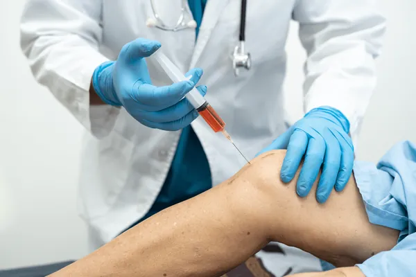
Understanding this anatomy explains why image-guided treatment matters. The intra-articular space is small and surrounded by major neurovascular structures (femoral artery and vein, femoral nerve, sciatic nerve). Blind landmark injection into the hip joint has published accuracy of only 50–65%, while fluoroscopic or ultrasound-guided injection is consistently >90% accurate. Treatment delivered into the wrong tissue does not work.
Hip Osteoarthritis vs. The Other Causes of Hip and Groin Pain
The single most important step in evaluating hip pain is determining where the pain actually is and what is generating it. The differential diagnosis is wide and the wrong diagnosis leads to the wrong treatment.
Hip osteoarthritis typically produces deep anterior groin pain, mechanical (worse with weight-bearing, better with rest), with morning stiffness, restricted internal rotation on exam, and X-ray findings of joint space narrowing and osteophytes. The C-sign is classic — patients cup the hand around the lateral hip in a C-shape to localize the pain.
Greater trochanteric pain syndrome (hip bursitis / GTPS) produces lateral hip pain over the bony prominence on the outside of the upper thigh, worse lying on the affected side at night, with tenderness on direct palpation. This is gluteus medius and minimus tendinopathy with secondary bursal inflammation — Modal Pain Management treats it on a separate page.
Sacroiliac (SI) joint dysfunction produces deep buttock pain just inferior and medial to the dimples of Venus (the Fortin finger sign), worse with single-leg standing and asymmetric loading, with positive provocative tests (FABER, distraction, compression, thigh thrust). Frequently mislabeled as lumbar disc or hip pain — in 15–30% of chronic low back pain cases, the SI joint is the actual pain generator.
Piriformis syndrome produces deep buttock pain that worsens with prolonged sitting and may radiate down the back of the leg, mimicking sciatica. Reproduced by the FAIR test.
Femoroacetabular impingement (FAI) and labral tear produces anterior groin pain with mechanical clicking, catching, or locking, classically reproduced by the FADIR test (flexion, adduction, internal rotation), more common in younger active patients (20s–40s) than osteoarthritis. May coexist with early OA.
Iliopsoas tendinitis or bursitis produces anterior groin pain reproduced by resisted hip flexion and direct palpation over the iliopsoas tendon. Often misdiagnosed as hip OA in active middle-aged patients with normal X-rays.
Inflammatory arthritis (rheumatoid, psoriatic, ankylosing spondylitis, reactive) produces hip pain with morning stiffness lasting more than 60 minutes, sometimes bilateral, with elevated inflammatory markers (ESR, CRP) and additional joint involvement. Requires rheumatology co-management.
Avascular necrosis (AVN) of the femoral head produces sudden or rapidly progressive groin pain in patients with risk factors (corticosteroid use, alcohol use, sickle cell disease, prior hip trauma, lupus). MRI is the test of choice — X-ray is normal in early disease.
Inguinal or sports hernia, adductor tendinopathy, and lumbar L3–L4 radiculopathy can all refer pain to the anterior hip and groin and must be considered.
Septic arthritis, fracture, and metastatic malignancy are emergencies that must be ruled out in the right clinical context (acute severe pain, fever, history of cancer, inability to bear weight).
The diagnostic workup at Modal Pain Management addresses each of these in a structured order so the right diagnosis drives the right treatment.
How Hip Osteoarthritis Is Diagnosed
The diagnosis of hip osteoarthritis is made by combining a structured history, a focused physical examination, targeted imaging, and — when needed — a diagnostic intra-articular injection.
The history asks about pain location (anterior groin is most specific for hip OA), pain character (deep mechanical ache rather than sharp shock), mechanical triggers (weight-bearing, stairs, getting up from low chairs), morning stiffness duration (15–60 minutes for OA; >60 minutes raises concern for inflammatory arthritis), prior hip injury or surgery, family history of joint replacement, occupational and recreational loading, and red flags (acute onset without trauma, fever, weight loss, history of cancer).
The physical examination systematically tests range of motion (internal rotation is the most sensitive early finding — restricted long before flexion or extension), provocative tests (FADIR for impingement and labral pathology, FABER for hip and SI joint, Stinchfield resisted-hip-flexion for iliopsoas), gait analysis (antalgic gait, Trendelenburg sign), muscle strength (hip abductors, flexors, extensors), neurovascular exam, and palpation of the trochanter, SI joint, and lumbar spine to address the differential.
Imaging begins with weight-bearing AP pelvis and frog-lateral hip X-rays — the standard initial workup that demonstrates joint space narrowing, osteophytes, subchondral sclerosis, subchondral cysts, and the Kellgren-Lawrence grade. MRI is reserved for cases where the X-ray does not match the clinical picture, where labral pathology, AVN, or stress fracture is suspected, or where surgical planning requires soft-tissue detail. High-resolution musculoskeletal ultrasound at the bedside during the consultation visit is increasingly valuable — it visualizes effusion, synovitis, dynamic impingement, iliopsoas tendinopathy, and guides therapeutic injection.
When clinical and imaging findings are mixed (early X-ray changes plus differential considerations), a diagnostic image-guided intra-articular hip injection with local anesthetic (with or without corticosteroid) is the gold standard — substantial pain relief during the anesthetic window confirms the hip joint as the dominant pain generator. This step changes management for many patients with confusing pictures.
The Image-Guided Treatment Ladder for Hip Osteoarthritis
Modal Pain Management uses a structured, evidence-based, image-guided ladder for hip osteoarthritis that is tailored to your disease stage, functional impairment, prior treatments, and goals.
Step 1 — Foundation: weight management plus structured physical therapy. A 5–10% body weight reduction reliably reduces hip joint load and is associated with significant pain reduction and slower radiographic progression — this is one of the highest-yield interventions in OARSI and AAOS guidelines. Structured physical therapy emphasizes hip abductor and core strengthening (the gluteus medius and minimus are the workhorses of pelvic stability), targeted range-of-motion work (especially internal rotation), gait retraining, and aquatic exercise for patients who cannot tolerate land-based loading. This foundation continues at every subsequent step on the ladder.
Step 2 — Image-guided intra-articular corticosteroid injection for fast flare control and to confirm the hip joint as the pain generator. Performed under fluoroscopic or ultrasound guidance for >90% accuracy (versus 50–65% blind), the injection delivers a corticosteroid-anesthetic mixture into the intra-articular space. Onset of relief is typically 3–7 days and meaningful pain reduction lasts 6–12 weeks in 70–80% of correctly diagnosed patients. Limit two corticosteroid injections per 12-month period at the same site.
Step 3 — Viscosupplementation (hyaluronic acid) or platelet-rich plasma (PRP) for durable medium-term relief. Image-guided intra-articular hyaluronic acid (off-label in the hip — FDA-approved in the knee) provides 4–6 months of relief in a substantial subset of patients without steroid-related concerns. Image-guided intra-articular PRP has the strongest emerging evidence for medium-term durable relief in mild-to-moderate hip OA — head-to-head studies (Battaglia, Sánchez and colleagues) show PRP outperforms hyaluronic acid at 6 and 12 months in suitable candidates. PRP is typically delivered as a series of 2–3 injections spaced 2–4 weeks apart and repeated annually.
Step 4 — Image-guided radiofrequency ablation of the articular branches of the femoral and obturator nerves. For patients with moderate-to-advanced hip osteoarthritis who are not candidates for or are not yet ready for total hip replacement, image-guided RFA of the sensory articular branches that innervate the hip capsule (analogous to genicular nerve RFA for the knee) is an emerging non-surgical option for sustained pain relief. The procedure first uses diagnostic blocks to confirm the appropriate target, then thermal RFA to interrupt pain signaling for 6–12+ months. This step is particularly valuable for patients who want to delay surgery, who are surgical risk (cardiac, pulmonary, or hematologic), or who have a wait until they can be scheduled for elective replacement.
Step 5 — Surgical referral for total hip arthroplasty. Modal Pain Management refers patients to NYC-based orthopedic surgeons when severe daily pain limits function despite the steps above, when X-ray demonstrates Kellgren-Lawrence grade 3 or 4 disease, when night pain disrupts sleep, and when the patient is a reasonable surgical candidate. Modern total hip replacement is one of the most successful operations in medicine, with 95%+ 10-year implant survival and substantial pain and function gains in well-selected patients. The role of pain management is to make sure that decision is being made for the right reasons and at the right time — not by default.
In parallel, patients with red flags or features suggesting inflammatory arthritis are referred for rheumatology workup; patients with suspected AVN or labral pathology get appropriate MRI and orthopedic consultation; and patients with red flags suggesting septic arthritis, fracture, or malignancy go to the emergency department.
Physical Therapy and Self-Care for Hip Osteoarthritis
Image-guided injection works best when paired with a structured exercise program. The evidence base is consistent: hip OA patients who combine intra-articular injection with 6–12 weeks of targeted physical therapy have substantially better and longer-lasting pain reduction than those who get injection alone. The core elements:
- Hip abductor strengthening — clamshells, side-lying leg lifts, banded lateral walks, single-leg bridges. The gluteus medius and minimus are the primary stabilizers of the pelvis during single-leg stance and are weak in nearly every patient with hip OA.
- Core and hip extensor work — bridges, dead bugs, bird dogs, modified planks. Pelvic stability reduces the asymmetric loading that worsens hip arthritis.
- Range-of-motion and stretching — gentle hip flexor stretches, internal rotation work in supine, supervised yoga or Pilates with appropriate modifications.
- Aquatic exercise — pool walking and aquatic therapy unload the joint by 50–80% depending on water depth and are excellent for patients who cannot tolerate land-based work.
- Stationary cycling — low-impact cardiovascular conditioning that maintains hip range of motion without high joint load. Recumbent or upright bicycle, both work.
Self-care measures that consistently help: weight management (single most impactful), supportive footwear with cushioning, limiting deep flexion postures (low chairs, deep recliners, long airline flights), sleeping with a pillow between the knees on the unaffected side, walking poles during flares (offload the hip 20–30%), and avoiding sudden increases in walking volume. Modal Pain Management coordinates these elements at the consultation visit.
When Hip Pain Is an Emergency
Most hip osteoarthritis is managed in the outpatient setting on the timeline of weeks. Certain presentations are emergencies and warrant immediate evaluation in the emergency department, not in the office:
- Acute severe hip pain with fever, chills, or recent infection — septic arthritis is a surgical emergency.
- Inability to bear weight on the leg after a fall or trauma — femoral neck or acetabular fracture must be ruled out, especially in older adults.
- Hip pain in a patient with a history of cancer — metastatic bone disease must be ruled out.
- Acute, severe groin pain in a patient on long-term corticosteroids, with sickle cell disease, or with heavy alcohol use — avascular necrosis of the femoral head can present this way and requires urgent MRI.
- New leg weakness, numbness, or loss of bowel or bladder control with hip and back pain — cauda equina syndrome requires emergency MRI and neurosurgical consultation.
If any of these apply, go to the emergency room first. Modal Pain Management is the right setting for non-emergency, structured hip osteoarthritis evaluation and treatment.
Why Modal Pain Management for Hip Osteoarthritis
Modal Pain Management treats hip osteoarthritis with the modern hip-preservation playbook described above — image-guided diagnosis, image-guided therapy, evidence-based stratification, structured physical therapy, and a clear referral pathway for surgical and rheumatology cases. Dr. Alex Movshis is a board-certified pain management physician based at 369 Lexington Avenue in Midtown Manhattan, with same-week consultations, most major insurance accepted, and image-guided procedures performed in-office under fluoroscopic or ultrasound guidance. Most patients leave the consultation visit with a clear diagnosis and a written treatment plan within 45 minutes, and most procedures are scheduled within 1–2 weeks. The goal is not to put off surgery for the sake of putting off surgery — it is to make sure that every patient with hip arthritis has had a fair shot at the high-quality non-surgical options that exist before going to the operating room.