Hip bursitis is one of the most common — and most misdiagnosed — sources of lateral hip pain in adults aged 40–70, especially women. At Modal Pain Management in Midtown Manhattan, Dr. Alex Movshis uses real-time ultrasound to image the trochanteric bursa, the gluteus medius and minimus tendons, and the iliotibial band — and to deliver image-guided injections precisely into the painful structure. Most patients walk out the same day with substantially less pain, return to work the next morning, and resume normal activities within one to two weeks.
This page covers what hip bursitis actually is (it is rarely just bursitis — it is usually a tendon problem with the modern name “greater trochanteric pain syndrome”), how it is distinguished from the other causes of lateral hip and buttock pain, how it is diagnosed at Modal Pain Management, and the evidence-based image-guided treatment ladder we use to get patients back to walking, sleeping on their side, climbing stairs, and exercising without pain.
Hip Bursitis Has Been Renamed: Greater Trochanteric Pain Syndrome (GTPS)
For decades the standard diagnosis for lateral hip pain was “trochanteric bursitis” — and the standard treatment was a cortisone injection into the bursa. Modern imaging changed that. When patients with classic “trochanteric bursitis” symptoms are evaluated with high-resolution ultrasound and MRI, only a minority have isolated bursal inflammation. The majority have tendinopathy of the gluteus medius and gluteus minimus tendons at their insertions on the greater trochanter — with the bursa inflamed secondarily because it sits superficial to those degenerated tendons.
Because the tendon is the primary pain generator in most cases, the umbrella diagnosis was renamed to greater trochanteric pain syndrome (GTPS). This terminology change has clinical consequences. A purely bursa-focused treatment plan (rest plus repeated cortisone injections) does not address — and can actually worsen — the underlying gluteal tendon disease. The image-guided ladder we use at Modal Pain Management explicitly accounts for the bursal versus tendinous components and selects treatments accordingly.
The four overlapping conditions that fall under the GTPS umbrella are:
- Gluteus medius tendinopathy — the most common, affecting the insertion on the lateral facet of the greater trochanter.
- Gluteus minimus tendinopathy — often coexists with gluteus medius involvement; affects the insertion on the anterior facet.
- Trochanteric bursitis (true bursal inflammation) — fluid distension of the bursa, often secondary to underlying tendinopathy.
- External snapping hip (coxa saltans externa) — the iliotibial band snapping over the greater trochanter with hip motion.
Distinguishing which of these is dominant — and treating accordingly — is what separates a successful 12-month outcome from a recurrence-prone “I keep getting cortisone shots that don’t last” pattern.
Why It Hurts on the Outside of Your Hip: The Anatomy
The greater trochanter is the bony prominence on the outer side of the upper thigh — easy to feel by pressing firmly on the lateral hip while lying on the opposite side. Several structures attach to or pass over the greater trochanter:
- Gluteus medius tendon inserts on the lateral and superoposterior facets of the trochanter; it is the primary hip abductor and the primary stabilizer of the pelvis on single-leg stance.
- Gluteus minimus tendon inserts on the anterior facet; it assists hip abduction and internal rotation.
- Trochanteric bursa is a fluid-filled sac that sits superficial to the gluteus medius tendon and deep to the iliotibial band, reducing friction during gait.
- Subgluteus medius and subgluteus minimus bursae sit deep to those tendons; they can also become inflamed.
- Iliotibial (IT) band passes directly over the greater trochanter as it travels from the iliac crest to the lateral knee.
The combination of compressive load (sleeping on the painful side, sitting cross-legged, single-leg stance with valgus collapse) plus repetitive friction from the IT band is what drives the gluteal tendinopathy and secondary bursal inflammation that we collectively call GTPS.
GTPS vs. Other Causes of Lateral Hip and Buttock Pain
Lateral hip pain has a long differential diagnosis, and the wrong diagnosis leads to predictable treatment failure. At Modal Pain Management we systematically rule out each of the following before settling on GTPS as the working diagnosis:
- Hip osteoarthritis (OA) — pain is typically in the groin, not the lateral hip; worse with internal rotation; positive FABER and FADIR tests; X-ray shows joint space narrowing. Some patients have both hip OA and GTPS simultaneously.
- Lumbar radiculopathy (L5 or S1) — pain radiates from the low back down the back or outer thigh past the knee; positive straight leg raise; weakness in dorsiflexion or plantarflexion; sensory deficits in dermatomal pattern. See our sciatica page.
- Piriformis syndrome — deep buttock pain that worsens with sitting and improves with walking; pain reproduced with the FAIR test; pain often radiates down the back of the thigh. See our dedicated piriformis syndrome page.
- Femoroacetabular impingement (FAI) and hip labral tear — younger active patients (20s–40s); groin pain with deep flexion, internal rotation, and pivoting; positive FADIR test; often confirmed on hip MR arthrogram.
- Sacroiliac (SI) joint dysfunction — pain over the SI joint and posterior pelvis; positive Patrick (FABER), thigh thrust, distraction, and compression tests; can refer to the lateral hip.
- Meralgia paresthetica — entrapment of the lateral femoral cutaneous nerve causing burning and numbness over the anterolateral thigh; classic “wallet pocket” or tight-belt-line distribution.
- Hip stress fracture (femoral neck or trochanter) — runners, military recruits, or patients with osteoporosis or relative energy deficiency; pain with weight-bearing; requires MRI to diagnose; missing this diagnosis can lead to a complete fracture.
- Septic bursitis or septic arthritis — warm, red, swollen hip with fever or chills; medical emergency requiring same-day aspiration and culture.
A focused physical exam plus bedside musculoskeletal ultrasound — both performed at the Modal Pain Management consultation — typically distinguishes GTPS from these mimickers in a single visit. X-rays are added when hip OA, fracture, or femoral pathology is suspected; MRI is reserved for atypical cases, suspected gluteal tendon tear requiring surgical evaluation, or refractory cases that have failed conservative treatment.
How GTPS Is Diagnosed at Modal Pain Management
Diagnosis at Modal Pain Management is anatomically specific from the first visit. Dr. Movshis performs a focused history (sleep position, sitting habits, recent activity changes, prior hip surgery, lumbar spine history, menopause status), a structured physical exam, and bedside musculoskeletal ultrasound during the consultation itself.
Key exam maneuvers include:
- Direct palpation over the greater trochanter, posterior facet (gluteus medius insertion), and anterior facet (gluteus minimus insertion) to identify the painful structure.
- Resisted hip abduction (in side-lying) and the single-leg stance test (a 30-second one-legged stand reproducing lateral hip pain is highly specific for gluteal tendinopathy).
- Trendelenburg test — pelvic drop on the contralateral side during single-leg stance indicates gluteus medius weakness or pain inhibition.
- FABER (Patrick) test — to screen for hip joint pathology and SI joint pain.
- FADIR test — to screen for femoroacetabular impingement and labral pathology.
- Straight leg raise and slump test — to rule out lumbar radiculopathy.
Bedside ultrasound directly visualizes:
- The trochanteric bursa — confirming or ruling out fluid distension.
- The gluteus medius and minimus tendons at their trochanteric insertions — assessing thickness, fibrillar pattern, partial-thickness tears, calcific deposits, and dynamic snapping with hip motion.
- The iliotibial band as it passes over the trochanter — identifying snapping hip and friction zones.
- The gluteus medius and minimus muscle bellies — screening for fatty atrophy, which suggests chronic full-thickness tendon tear and changes management.
This single-visit ultrasound assessment is the difference between treating GTPS and treating the wrong target. X-rays, MRI, and lumbar imaging are added only when indicated by the exam.
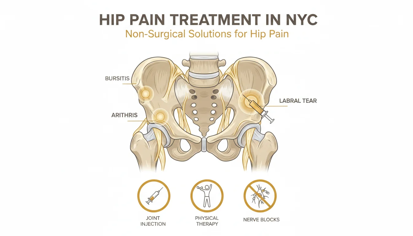
The Image-Guided Treatment Ladder for GTPS
Modal Pain Management treats GTPS using a stepwise, evidence-based ladder. Each step is selected based on the bursa-versus-tendon balance identified on ultrasound, the chronicity of symptoms, and how much functional improvement the patient needs.
Step 1 — Load Management and Physical Therapy (6–8 weeks)
For acute and subacute GTPS, the strongest evidence supports a structured, education-plus-exercise program. The 2018 LEAP randomized trial showed that 8 weeks of education plus loaded exercise produced higher rates of “much better” or “fully recovered” outcomes than either corticosteroid injection or watch-and-wait — and the advantage persisted at 12 months. The program includes:
- Postural education: stop sleeping on the painful side (use a pillow between knees or sleep on the opposite side); stop sitting cross-legged; stop standing with weight shifted onto one hip; stop crossing the legs at the knees.
- Isometric gluteal loading: side-lying hip abduction holds, banded clamshells, isometric wall press of the abducted leg.
- Progressive isotonic loading: side-lying hip abduction, banded side-stepping, single-leg bridges, step-ups.
- Eccentric and functional progression: lateral lunges, single-leg deadlifts, step-downs, gait retraining to reduce pelvic drop and valgus knee collapse.
- Treatment of contributing biomechanics: addressing leg-length discrepancy with a heel lift, treating co-existing lumbar pathology, and screening for total hip replacement-related lateral mechanics.
Physical therapy is foundational — patients who skip this step are the patients who recur.
Step 2 — Ultrasound-Guided Corticosteroid Injection
For patients with substantial bursal inflammation on ultrasound, severe pain limiting their ability to sleep or work, or those who need fast functional improvement for an upcoming event, ultrasound-guided trochanteric bursa corticosteroid injection is added on top of the exercise program — not as a replacement for it. Image guidance matters: blind landmark injections into the trochanteric bursa miss the target in 30–40% of cases, leading to underwhelming relief. Ultrasound guidance pushes accuracy to 95%+, allows safe avoidance of the gluteal tendon and the sciatic nerve posteriorly, and is the standard of care for every bursa procedure at Modal Pain Management.
Substantial pain relief typically begins 3–7 days after injection and continues to improve over 2–4 weeks. We limit corticosteroid injections to no more than two per 12-month period at the same site — repeated steroid exposure carries a dose-dependent risk of weakening the gluteus medius and minimus tendons, which is the opposite of the long-term goal in GTPS.
Step 3 — Ultrasound-Guided PRP (Platelet-Rich Plasma) Injection
For chronic GTPS (>3 months), patients with imaging evidence of gluteal tendinopathy or partial-thickness tears, patients who have already received one or two cortisone injections, and patients trying to avoid further tendon injury, we recommend ultrasound-guided PRP injection of the gluteus medius and minimus tendons. PRP works by delivering a concentrated dose of the patient’s own platelet-derived growth factors directly into the degenerated tendon, stimulating regenerative healing rather than simply suppressing inflammation.
Recent randomized controlled trials (Begkas et al., 2020; Fitzpatrick et al., 2018, 2019 with 2-year follow-up) consistently show that for chronic gluteal tendinopathy, PRP outperforms repeat corticosteroid injection at 12-month and 2-year endpoints. The clinical effect is slower (maturation over 6–12 weeks) but more durable, and PRP avoids the tendon-weakening risk of repeated steroid. At Modal Pain Management, PRP is performed under continuous ultrasound guidance with peppering of the affected tendon segments, followed by a structured 6-week loading progression.
Step 4 — ESWT and Surgical Referral (Selected Cases)
For refractory cases that have failed steps 1–3, options include:
- Extracorporeal shockwave therapy (ESWT) — focused or radial shockwave applied to the gluteal tendons; we coordinate referral to NYC providers offering this modality.
- Surgical referral — reserved for patients with full-thickness gluteus medius or minimus tendon tears confirmed on MRI, particularly those with persistent abductor weakness, marked Trendelenburg gait, or post-total-hip-replacement abductor mechanism failure. We refer to selected NYC orthopedic hip specialists for endoscopic or open gluteal tendon repair.
The vast majority of patients (>90%) achieve durable relief without ever reaching step 4.
Physical Therapy and Self-Care for GTPS
Even the best image-guided injection is only as durable as the postural and biomechanical work that follows. Patients with GTPS benefit from a structured course of physical therapy targeting:
- Hip abductor strength — particularly the posterior fibers of the gluteus medius, which control pelvic drop on single-leg stance.
- Gluteal endurance — graduated loading to build the capacity to withstand walking, stair climbing, and prolonged standing without symptoms.
- Lumbopelvic stability — core and hip control work to address the pelvic drop / valgus knee collapse pattern.
- Calf, hamstring, and IT band flexibility — reducing the friction load on the trochanteric structures.
- Gait retraining — for runners, addressing overstriding, cadence, hill running volume, and shoe wear.
Home self-care includes:
- Sleep position modification — sleep on the unaffected side with a thick pillow between the knees to keep the upper hip in neutral abduction; if you must sleep on the painful side, use a softer mattress or memory-foam topper.
- Sitting position modification — uncross the legs; sit with both feet flat on the floor; rise from low chairs using both legs symmetrically.
- Standing posture — distribute weight evenly between both feet; avoid “hanging on the hip” with weight shifted to one side.
- Activity modification — temporarily reduce hill walking, treadmill incline work, and high-volume stair climbing; resume gradually as symptoms allow.
- NSAIDs — naproxen 220–440 mg twice daily or ibuprofen 400–600 mg three times daily for 7–14 days during flares, unless contraindicated.
When Hip Pain Is an Emergency
Most lateral hip pain is not an emergency — but a few presentations require same-day evaluation:
- Septic bursitis or septic arthritis — warm, red, markedly tender hip with fever, chills, or recent overlying skin infection. Diabetic, immunosuppressed, and recent-injection patients are at highest risk. Aspiration with culture is required before any steroid is administered.
- Acute trauma with inability to bear weight — particularly in older patients or those with osteoporosis. Hip fracture must be ruled out with X-ray (and sometimes MRI for occult fractures).
- Stress fracture in a runner — progressive hip pain with weight-bearing in a high-mileage runner, military recruit, or patient with relative energy deficiency. MRI is required for diagnosis. A missed femoral neck stress fracture can progress to a complete, displaced fracture.
If any of these descriptions fit, go to an emergency room or call our office urgently rather than scheduling a routine consultation.
Why Modal Pain Management for Hip Bursitis and GTPS in NYC
Modal Pain Management is a physician-led interventional pain practice located at 369 Lexington Avenue, Floor 25 in Midtown Manhattan (NYC 10017), led by Dr. Alex Movshis, a dual board-certified pain management physician fellowship-trained at the Icahn School of Medicine at Mount Sinai. Every patient is evaluated and treated by Dr. Movshis personally — not by a rotating cast of providers. Bedside musculoskeletal ultrasound is available at the consultation visit, which means most hip bursitis and GTPS cases are diagnosed and have a treatment plan in place at a single appointment. Same-week new-patient appointments are typically available.
For sourced clinical evidence on the procedures referenced on this page — including efficacy ranges, recovery timelines, and PRP-vs-corticosteroid comparison data — see the clinical evidence and citations page. Most commercial PPO insurance plans are accepted; Medicare and Medicaid are not. Insurance benefits are verified before your visit at no charge or obligation through the insurance verification form.