At Modal Pain Management in Midtown Manhattan, Dr. Alex Movshis performs image-guided sacroiliac (SI) joint injections as both the diagnostic gold standard and the first-line therapeutic option for one of the most commonly missed causes of chronic low back pain. Sacroiliac joint dysfunction is responsible for an estimated 15–30% of chronic low back pain cases — yet lumbar MRI almost always looks unremarkable at the SI joint, the pain pattern overlaps with lumbar disc, facet, and hip disease, and the diagnosis is frequently delayed by months or years. Image-guided SI joint injection — performed under live fluoroscopy or ultrasound, with contrast confirmation of intra-articular placement — is the only reliable way to both confirm the SI joint as the dominant pain generator and deliver therapeutic anti-inflammatory medication directly into the joint capsule. Located at 369 Lexington Avenue Floor 25 in NYC 10017, we perform SI joint injections using the technique and evidence base endorsed by the American Society of Regional Anesthesia (ASRA), the Spine Intervention Society (SIS), and the 2020 ASIPP guidelines.
What the Sacroiliac Joint Is — and Why It Hurts
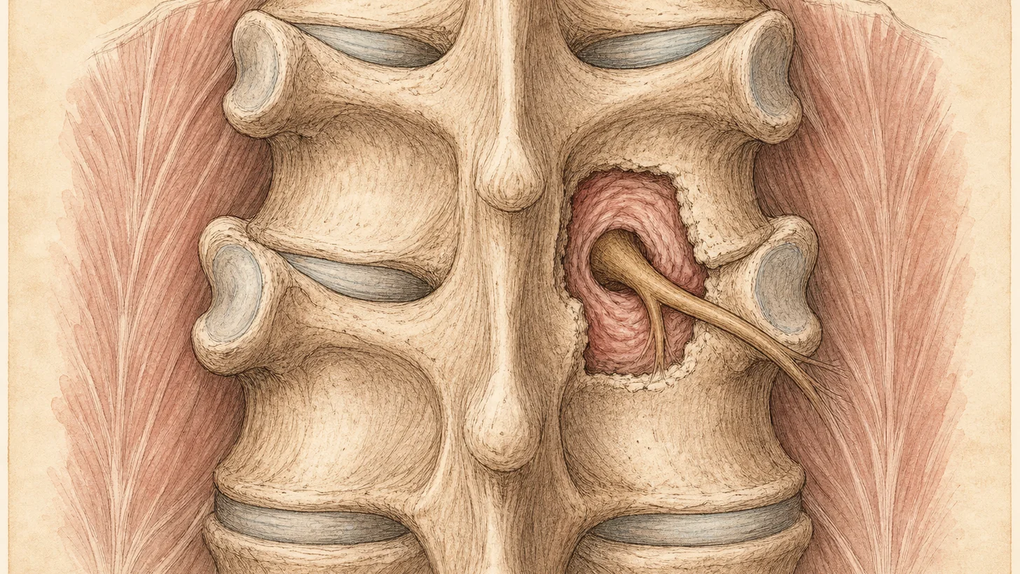
The sacroiliac (SI) joint is the junction between the sacrum (the triangular bone at the base of the spine, formed by the fusion of the lower five vertebrae) and the ilium (the wide, flat bone that forms the upper part of the pelvis). There is one SI joint on each side. The joint is small, powerful, and designed primarily for load transfer: it transmits force between the spine and the lower limbs and absorbs the shock of standing, walking, and running. The joint surfaces are irregular and interlocking, stabilized by one of the densest networks of ligaments in the body (the anterior sacroiliac, interosseous, dorsal sacroiliac, sacrotuberous, and sacrospinous ligaments). The joint capsule is richly innervated by the lateral branches of the L5 dorsal ramus and the S1, S2, and S3 dorsal sacral rami — the small sensory nerves that carry SI joint pain to the spinal cord and brain.
SI joint pain arises from three overlapping pathophysiologies. Mechanical dysfunction — a subtle abnormality of SI joint motion, often related to pregnancy, trauma (a fall on the buttock, a motor vehicle accident, a single heavy lift with rotation), pelvic asymmetry, or compensatory motion after lumbar spine fusion surgery — is the most common. Inflammatory sacroiliitis — true inflammation of the SI joint driven by an underlying systemic inflammatory arthritis such as ankylosing spondylitis, psoriatic arthritis, or inflammatory bowel disease-associated spondyloarthritis — is less common but clinically important to recognize because it changes the entire treatment plan (patients need disease-modifying systemic therapy, not just local injections). Post-traumatic and post-surgical instability — a looser, hypermobile SI joint following pelvic trauma or lumbosacral fusion — is a third pattern, sometimes requiring structural stabilization in addition to interventional pain management. Image-guided SI joint injection is appropriate for all three patterns, though the long-term strategy differs.
The clinical presentation is distinctive once recognized. SI joint pain is typically one-sided, centered over the posterior superior iliac spine (the bony prominence roughly two inches lateral to the base of the spine) and the upper buttock, and may refer into the groin, the lateral thigh, or the posterior thigh down to the knee. It rarely refers below the knee — which is a useful clinical discriminator versus lumbar radiculopathy. The pain is worse with prolonged sitting (particularly on hard surfaces), worse when standing up from a chair or car, worse rolling over in bed, and worse with stairs or single-leg weight-bearing. Patients often unconsciously shift their weight to the unaffected side when standing.
Clinical Evaluation — How We Confirm the SI Joint Before Injecting
At the consultation visit, Dr. Movshis performs a structured clinical evaluation designed to determine whether the SI joint is the dominant pain source, or whether the lumbar spine, the hip, or another structure is the primary driver. The evaluation uses the Laslett cluster of provocation tests — a validated battery of six clinical maneuvers first described by Mark Laslett and colleagues and since confirmed in multiple prospective studies. The six tests are:
The thigh thrust test (also called the femoral shear test) — with the patient supine and the hip flexed to 90°, a downward force is applied through the long axis of the femur, loading the SI joint through the femoral head.
The distraction test — with the patient supine, posteriorly directed force is applied to the anterior superior iliac spines bilaterally, stressing the anterior sacroiliac ligaments.
The compression test — with the patient in side-lying position, a downward force is applied to the iliac crest, stressing the dorsal sacroiliac and interosseous ligaments.
The sacral thrust test — with the patient prone, a downward force is applied to the center of the sacrum, stressing the joint bilaterally.
The Gaenslen’s test — with the patient supine near the edge of the exam table, one hip is fully flexed and the other is passively extended off the table, stressing both SI joints in opposite directions.
The FABER (Patrick) test — with the patient supine, the hip is Flexed, ABducted, and Externally Rotated in a figure-four position, with downward pressure on the flexed knee.
A positive Laslett cluster — three or more of the six tests reproducing the patient’s typical pain — has a sensitivity of approximately 91% and a specificity of approximately 78% for SI joint pain in the published literature (Laslett 2005, van der Wurff 2006). A normal lumbar MRI, a hip MRI that rules out intra-articular hip pathology, and the absence of true below-the-knee radicular symptoms strengthen the clinical picture.
Imaging is also reviewed. Plain pelvic X-rays and CT evaluate for sacroiliitis (erosions, sclerosis, joint space narrowing — seen in ankylosing spondylitis and other inflammatory arthropathies), pelvic asymmetry, and prior lumbosacral fusion. MRI of the SI joints with STIR sequences can reveal active inflammation (subchondral bone marrow edema) in inflammatory sacroiliitis. Laboratory workup — including HLA-B27, ESR, and CRP — is indicated when inflammatory arthropathy is suspected.
How the Procedure Works
The image-guided SI joint injection is performed in our in-office procedure suite under sterile conditions. The patient is positioned prone (face down) with a small pillow under the pelvis to flatten the lumbar spine and improve access to the SI joint. The skin over the posterior superior iliac spine and the lower SI joint is cleaned with chlorhexidine or povidone-iodine, and the surrounding skin is draped. A small amount of lidocaine is injected into the skin and subcutaneous tissue over the planned needle entry point — this initial numbing is the only meaningful sting most patients feel.
Under live fluoroscopic guidance — the gold-standard image-guidance technique for intra-articular SI joint injection — a small-gauge procedural needle (typically 22 gauge, 3.5 inches) is advanced through the posterior approach toward the inferior third of the SI joint. The inferior third is anatomically the portion of the joint most accessible to direct needle entry; the upper two-thirds of the joint are too posteriorly oriented to be reached through a standard posterior approach. Once the needle tip is positioned at the joint edge, a small amount of iodinated contrast (0.5–1 mL) is injected under live fluoroscopy. Confirmation of intra-articular spread — the characteristic arthrogram that outlines the curved, irregular contour of the SI joint space — is the critical safety check that confirms correct placement before the therapeutic injectate is delivered. Studies going back to Pekkafahli 2003 and Rosenberg 1999 have demonstrated that blind (non-image-guided) SI joint injections miss the joint in 60–78% of attempts — which is why image guidance is considered the standard of care for any diagnostic or therapeutic intra-articular SI joint injection.
Once intra-articular placement is confirmed, the therapeutic injectate — typically 1–2 mL of 0.5% bupivacaine (long-acting local anesthetic) mixed with 40 mg of triamcinolone or an equivalent long-acting corticosteroid — is slowly injected. The local anesthetic provides the diagnostic signal (the 4–24 hour anesthetic window during which pain reduction is measured); the corticosteroid provides the therapeutic effect (anti-inflammatory, taking 3–7 days to begin and 2–3 weeks to peak).
Ultrasound-guided SI joint injection is an acceptable alternative in selected cases, particularly for periarticular techniques that target the dorsal sacroiliac ligaments and the interosseous ligament rather than the intra-articular space. Ultrasound avoids radiation exposure (an important consideration in pregnant patients and in younger patients with a lifetime interventional trajectory) but cannot confirm intra-articular placement with the same certainty as fluoroscopy with contrast. Modal Pain selects the imaging modality based on the clinical target, the patient’s anatomy, and the published evidence for each specific indication.
The total procedure time is 15–25 minutes from skin prep to injection completion. After the injection, the patient is observed for 15–20 minutes in the recovery area, given a structured pain diary, and discharged home with post-procedure instructions.
The Evidence Base
The evidence base supporting image-guided sacroiliac joint injection is well-established across multiple decades of interventional pain research. The foundational epidemiologic studies by Schwarzer et al. 1995 and Maigne et al. 1996 established that the SI joint is the dominant pain generator in approximately 15–30% of patients presenting with chronic low back pain — a figure confirmed in subsequent prospective cohort studies. These studies used double-block validation (two separate injections with different anesthetic agents producing concordant relief) as the reference standard, and remain the basis for current clinical practice.
Therapeutic effectiveness of image-guided corticosteroid SI joint injection has been established in multiple prospective studies and systematic reviews. The Cohen 2005 comprehensive review published in Anesthesia & Analgesia summarized the accumulated evidence and established the framework of diagnostic-plus-therapeutic SI joint injection that remains standard today. The Simopoulos 2020 systematic review and ASIPP guidelines — the most recent comprehensive evidence synthesis — graded image-guided SI joint injection as a Level II recommendation with moderate evidence for short-term (≤12 week) pain relief in correctly selected patients, with 50–80% of patients achieving meaningful pain reduction for 6–12 weeks per injection.
The accuracy of image-guided versus blind technique is settled. Pekkafahli 2003 demonstrated that fluoroscopically-guided injection achieves intra-articular placement in 90%+ of attempts, versus approximately 22% for blind injection. Rosenberg 1999 demonstrated essentially the same result using post-injection arthrography. The Bollow 1996 and Braun 1996 MRI-verification studies added further support. These findings are the reason modern interventional pain guidelines consider image guidance the standard of care for any diagnostic or therapeutic SI joint injection.
For patients who require more durable relief than corticosteroid provides, the evidence base for lateral branch radiofrequency ablation of the L5 dorsal ramus and the S1–S3 lateral branches has grown substantially over the last decade. Cohen 2008, Patel 2012 (randomized controlled trial), Hansen 2012, and multiple subsequent meta-analyses have established that image-guided cooled-tip lateral branch RFA produces 6–12 months of clinically meaningful pain reduction in correctly selected patients — a durability that meaningfully exceeds what steroid alone provides.
Who Is a Good Candidate — and Who Isn’t
Image-guided SI joint injection is the right procedure for several specific clinical scenarios and the wrong procedure for others. The consultation visit produces a clear yes/no/with-modifications answer for each patient.
The strongest candidates are: patients with chronic SI joint pain lasting 6–12 weeks or longer who have not responded to conservative measures (physical therapy, oral anti-inflammatories, activity modification, supportive belt); patients with a clinical picture consistent with SI joint pain — unilateral posterior superior iliac spine and buttock pain, a positive Laslett cluster, no below-the-knee radicular symptoms, normal or unremarkable lumbar MRI; patients with post-pregnancy SI joint dysfunction (extremely common, often missed in primary care, highly responsive to image-guided injection plus targeted pelvic floor and gluteal physical therapy); patients with post-lumbar-fusion SI joint pain (the adjacent-segment SI joint is mechanically overloaded after L4–L5 or L5–S1 fusion and is a well-recognized cause of persistent post-surgical back pain); and patients with inflammatory sacroiliitis in whom the injection provides symptomatic control while systemic disease-modifying therapy is being optimized.
The procedure is not the right first-line treatment for: acute low back pain within the first 6 weeks (conservative care is appropriate first); pain that is primarily below the knee (true lumbar radiculopathy is more likely the source, and lumbar epidural steroid injection is the better option); pain with red-flag features (fever, weight loss, progressive neurologic deficit, saddle anesthesia, bladder/bowel dysfunction — these require urgent workup and often surgical consultation, not interventional pain management); suspected septic sacroiliitis (a medical emergency); active local skin infection over the injection site; and patients on uncontrolled anticoagulation.
Comparison vs. Other Low-Back Pain Treatments
Patients evaluating their options for chronic low back and buttock pain frequently want a side-by-side comparison of where SI joint injection fits on the broader treatment ladder.
Compared to lumbar epidural steroid injection: the two procedures target fundamentally different pain sources — lumbar ESI treats nerve-root pain from disc herniation, spinal stenosis, or lumbar radiculitis, while SI joint injection treats joint capsule and ligamentous pain from SI joint dysfunction. Patients with below-the-knee radicular symptoms, a positive straight-leg raise, and lumbar MRI showing nerve-root compression are lumbar ESI candidates. Patients with unilateral posterior superior iliac spine pain, a positive Laslett cluster, and unremarkable lumbar MRI are SI joint injection candidates. A significant number of patients have both — mixed lumbar and SI joint contributions — and a structured diagnostic workup (selective injections to each potential source) is the most reliable way to sort it out.
Compared to lumbar facet (medial branch) block: both target posterior-element low-back pain, but the anatomic targets are different. Lumbar medial branch blocks target the facet joints of the lumbar spine — typically L3–L4, L4–L5, and L5–S1 — and the pain pattern is usually midline or paramedian, worse with extension and rotation. SI joint pain is one-sided, centered over the posterior superior iliac spine and upper buttock, and worse with sitting and transitional movement (standing up from a chair). Patients with mixed facet and SI joint pain often benefit from sequential workup of each.
Compared to lateral branch radiofrequency ablation of the SI joint: the injection is faster-onset (3–7 days to meaningful relief versus 2–6 weeks for RFA) and appropriate for first-line and occasional use. The RFA is slower-onset but longer-lasting (6–12 months versus 6–12 weeks) and appropriate for patients who have confirmed the SI joint as the pain source and need a more durable option. The two procedures are sequential phases of the same treatment ladder, not competitors.
Compared to surgical SI joint fusion: surgical SI joint fusion — minimally invasive or open — is a definitive procedure that eliminates SI joint motion by fusing the sacrum to the ilium with titanium implants. It is appropriate for patients with severe, refractory SI joint pain that has failed all conservative and interventional options including repeat injection and lateral branch RFA, and who have true mechanical instability on imaging. The procedure carries surgical risk (infection, implant failure, adjacent-segment disease), is irreversible, and has a 6–12 week recovery. Modal Pain Management works with orthopedic and neurosurgical spine colleagues at NYU Langone and other NYC centers when surgical fusion becomes the appropriate option; the majority of our SI joint patients are effectively managed with image-guided injection alone or in combination with lateral branch RFA, without surgery.
Compared to physical therapy alone: structured physical therapy targeting the gluteus medius, the core, the pelvic floor, and the hip abductors is the foundation of SI joint management and should be ongoing regardless of what interventional procedures are performed. Injection and PT are not competitors — they are complementary. The injection reduces pain to a level at which productive PT becomes possible; the PT addresses the underlying mechanical dysfunction that drives the pain. Patients who rely on injection alone without addressing the mechanical contributors tend to require more frequent injections; patients who combine injection with sustained PT tend to get longer durations of relief per injection and fewer injections per year.
What to Expect: The Patient Pathway
The full pathway from initial consultation to therapeutic SI joint injection at Modal Pain Management typically runs 2 to 4 weeks for a new patient.
Initial consultation visit (45 minutes). Dr. Movshis reviews your back and buttock pain history, prior imaging, prior treatments and responses, and overall medical context. A structured physical examination — including the full Laslett cluster, a lumbar spine exam, a hip exam, and a neurologic exam — sorts out the dominant pain generator and rules out referred pain. Imaging is reviewed. The diagnostic plan, the indications for SI joint injection, the expected response rates, the alternatives, and the cost and insurance picture are discussed in plain language. Most patients leave the consultation with a clear written plan.
Insurance authorization (5–10 business days). Our team submits the prior authorization request with documentation of conservative care, examination findings, and imaging. Most commercial PPO plans approve image-guided SI joint injection when the documentation supports the indication.
Image-guided SI joint injection (15–25 minute procedure). Performed in our Midtown NYC office under live fluoroscopic or ultrasound guidance, with no IV sedation in most cases. You drive home after a 15–20 minute observation period and record your pain in a structured diary every 30–60 minutes for 4–8 hours after the injection.
Follow-up visit (15 minutes, 2–3 weeks after the injection). The pain diary is reviewed and the response objectively measured. Diagnostic signal (the 4–24 hour anesthetic window) is interpreted; therapeutic response (the 2–3 week steroid effect) is assessed. The long-term plan is updated — repeat injection on schedule, progression to lateral branch RFA, or escalation to surgical consultation — based on the clinical picture.
Ongoing management. Most patients transition to a structured gluteal/core physical therapy program after the injection, return for a repeat injection if and when pain recurs, and escalate to RFA if they find themselves needing more than 2–3 injections per year.
Recovery, Activity, and Return to Function
Recovery from image-guided SI joint injection is rapid. Post-procedure soreness at the injection site is common for 2–4 days and managed with ice and over-the-counter analgesics. A brief 1–3 day post-procedure pain flare — a temporary increase in pain before the steroid begins to work — occurs in 5–10% of patients and is self-limited. Most patients take the day of the procedure off, resume desk-based work the following day, and resume light walking the same evening.
Activity guidance for the first 2 weeks includes: avoiding high-impact loading (running, jumping, heavy resistance work), avoiding prolonged sitting on hard surfaces when possible, and beginning or continuing a gluteal-medius and core stabilization program. Most patients note meaningful pain improvement by 7–14 days as the steroid effect takes hold, and full therapeutic benefit at 3 weeks. A follow-up visit at 2–3 weeks objectively measures the response and sets the long-term plan.
Driving is permitted as soon as the patient is comfortable — typically the same day. Return to full unrestricted activity including recreational sports is typically possible by 2–3 weeks.
Insurance, Authorization, and Practical Logistics
Most commercial PPO insurance plans cover image-guided sacroiliac joint injections when ordered as part of a structured workup for chronic low back or buttock pain. Coverage typically requires prior authorization documenting 6–12 weeks of conservative care, a clinical examination consistent with SI joint pain, and an appropriate differential workup. We handle the prior authorization process on your behalf.
We accept most major commercial PPO plans (United Healthcare, Aetna, Cigna, BlueCross BlueShield, Oxford, Empire BCBS) and do not accept Medicare, Medicaid, HMO plans, or workers’ compensation. Out-of-pocket cost depends on your individual deductible and coinsurance — we verify benefits and provide a written estimate before the procedure. Visit our insurance verification page to check your plan, or call (646) 290-6660 for a benefits check.
For patients without insurance coverage or for whom out-of-network self-pay is the right option, transparent self-pay pricing is provided at the consultation.
Why Patients Choose Modal Pain Management for SI Joint Injections
Dr. Alex Movshis is board-certified in Anesthesiology with subspecialty fellowship training in Interventional Pain Medicine, and performs all sacroiliac joint injections under live fluoroscopic or ultrasound guidance with contrast confirmation — the technique standard endorsed by ASRA, the Spine Intervention Society, and ASIPP. We treat the SI joint injection as a structured diagnostic and therapeutic tool, not a one-off intervention: every injection is paired with a documented pain diary, a follow-up visit to objectively measure response, and a clear long-term plan that escalates to lateral branch radiofrequency ablation or refers to surgical colleagues when appropriate.
Our Midtown NYC office at 369 Lexington Avenue Floor 25 is one block from Grand Central Terminal, making it accessible from anywhere in the New York metro area. Same-week consultation appointments for new patients are typically available.
If you have chronic low back or buttock pain that has not responded to physical therapy and conservative care, unilateral pain centered over the posterior superior iliac spine, pain that is worse with sitting and standing up from a chair, or pain after pregnancy or lumbar fusion surgery that fits the SI joint pattern — image-guided SI joint injection may be the right next step. Learn more about the underlying condition on our SI joint pain page, or book a consultation to discuss whether you are a candidate.