Roughly 10–15% of patients who have a major surgery develop chronic nerve pain that persists beyond the expected healing window (>3 months from the index operation defines chronic post-surgical pain in the IASP literature). For a small subset of surgeries — open thoracotomy, mastectomy with axillary dissection, certain hernia repairs — the rate climbs above 25%. The pain is not in the patient’s head, it is not “just scar tissue,” and it is almost never solved by another operation in the same field.
Most of these cases share a specific peripheral nerve generator that can be identified, confirmed with an image-guided diagnostic block, and treated directly. The clinical work is matching the patient’s pain pattern to the responsible nerve, confirming the diagnosis with a 10–15 minute ultrasound-guided block, and walking through a procedural ladder that escalates only as needed.
This is one of the most under-served niches in interventional pain medicine. Surgeons rarely follow patients into chronic post-surgical pain because their training focus is the operation itself, neurologists treat the medication side, and most pain clinics see iatrogenic neuropathies infrequently enough that the relevant blocks are not part of their routine offerings. Modal Pain Management is built around this work. Dr. Alex Movshis is dual board-certified in Anesthesiology and Pain Medicine and fellowship-trained at the Icahn School of Medicine at Mount Sinai, with a clinical focus on post-surgical neuropathic pain — same-week new-patient consultations are routinely available at 369 Lexington Avenue Floor 25 in Midtown Manhattan.
Chronic pain after specific surgeries
Inguinal and ventral hernia repair
The clinical pattern is anchored by PAT-001 above. The three nerves at risk are the ilioinguinal, iliohypogastric, and genitofemoral. Open Lichtenstein repair classically injures the ilioinguinal nerve at the external oblique aponeurosis, often by direct ligation, suture entrapment, or mesh contact. Laparoscopic TEP/TAPP repair places the genitofemoral nerve at risk where the mesh sits over the iliopubic tract. Ventral hernia and incisional hernia repairs put the lower intercostal cutaneous branches (T10–T12 anterior cutaneous) at risk in the same way ACNES is generated by laparoscopic port placement.
The pain typically begins within the first six weeks after surgery and either fails to resolve or worsens past the 12-week point. It is burning, lancinating, or “electric” in quality, often with a Tinel-positive point along or just lateral to the incision. The diagnostic workup is sequential image-guided blocks — ilioinguinal first for open repairs, genitofemoral first for laparoscopic mesh repairs, and so on. A positive block (≥50% pain abolition in the working window) identifies the dominant nerve and, with steroid added, produces 4–12 weeks of therapeutic relief. Patients with clear diagnostic response and diminishing duration are candidates for pulsed RFA. Refractory cases are referred for peripheral-nerve surgical evaluation — the Amid triple neurectomy, performed by a peripheral nerve plastic surgeon, is the gold-standard salvage operation for inguinodynia.
C-section and Pfannenstiel incisions
Hysterectomy and pelvic surgery
Many patients with this pattern have been dismissed for years as having “adhesions,” “recurrent endometriosis,” or “pelvic floor dysfunction” — and have been on hormonal suppression and pelvic-floor PT without meaningful change. The diagnostic block is the test their workup is missing. See the dedicated post on chronic pelvic pain after hysterectomy for the full nerve-by-nerve workflow.
Total hip replacement
Total hip arthroplasty involves three distinct neuropathic risks. The lateral femoral cutaneous nerve is at risk during anterior approach THA — the same nerve that produces meralgia paresthetica in non-surgical patients (see the entrapment page). The obturator nerve can be irritated by acetabular cup positioning or by extra-articular cement extrusion, producing deep groin and medial-thigh pain. The sciatic nerve is rarely transected but can be stretched during posterior approach exposure, producing posterior-thigh or calf neuropathic pain.
Anterior approach THA is the most common modern technique and meralgia paresthetica is the most common iatrogenic neuropathy after it — typically presenting 4–12 weeks after surgery with the classic anterolateral thigh burning pattern. Diagnostic ultrasound-guided LFCN block confirms the diagnosis in 10–15 minutes. The hip implant is rarely the problem in these cases.
Mastectomy and axillary dissection
Thoracotomy and rib surgery
Approximately 43% of open thoracotomy patients and 20–35% of VATS patients develop chronic post-thoracotomy pain syndrome (PTPS). The mechanism is intercostal nerve injury at the rib retraction sites (open) or trocar entries (VATS). The clinical pattern divides into neuropathic intercostal pain (burning, electric, dermatomal, allodynic) and mechanical chest-wall pain (deep, exertional, reproduced by twisting or coughing). Most patients have a mixed picture. Diagnostic ultrasound-guided intercostal nerve blocks at the affected levels separate the components and identify the dominant target. See the dedicated post-thoracotomy pain syndrome page for the full ladder including paravertebral blocks, intercostal pulsed RFA, and cryoablation.
Spine surgery and failed back surgery syndrome
The umbrella term “failed back surgery syndrome” combines five mechanistically distinct entities, and treating them as a single diagnosis is the most common reason patients fail conservative care. Adjacent-segment disease (degenerative change at the level above or below a fusion) presents as recurrent radicular pain from a new disc or foraminal stenosis. Epidural fibrosis produces persistent radicular pain from scar tethering the nerve root — diagnosed by MRI with contrast. Adjacent-segment sacroiliac dysfunction — 40–43% of fusion patients develop SI joint pain within 5 years — presents as buttock/posterior-thigh pain and is diagnosable with an SI joint block. Cluneal nerve entrapment at the iliac crest is a frequent miss after spine surgery with iliac crest bone graft harvest. Painful neuroma at the surgical scar — particularly the medial branch of the dorsal ramus — produces focal Tinel-positive pain along the incision. Each has a different diagnostic anchor and a different procedural answer; the diagnostic block sequence sorts them out.
What scar tissue actually does to nerves
The lay phrase “scar tissue pain” obscures three distinct pathological mechanisms, each with different treatment implications.
Perineural fibrosis. Connective-tissue scar forms around an intact nerve, mechanically tethering it to surrounding tissue. The nerve loses its normal gliding excursion through the tissue plane and develops symptoms with movement or limb positioning. Mechanism: dense collagenous fibrosis without breach of the nerve’s epineural sheath. Treatment: ultrasound-guided hydrodissection — injection of saline, 5% dextrose, or dilute steroid into the fascial plane around the nerve mechanically separates the perineural scar and restores nerve excursion. Often dramatic in benefit; commonly repeatable.
Neuroma-in-continuity. Intra-fascicular injury without complete transection. The nerve remains in continuity but a focal segment becomes hypersensitive — pathologically firing in response to normal mechanical loading and to mild stretching. Imaging may show focal thickening on high-resolution ultrasound or MR neurography. Treatment ladder: pulsed RFA → cryoneurolysis → surgical neurolysis with peripheral nerve plastic surgery.
Terminal neuroma. The nerve was cut. The proximal stump forms a disorganized ball of regenerating axons trying to find their distal target. Terminal neuromas are typically Tinel-positive, palpable, and exquisitely tender at a discrete point. Treatment: percutaneous radiofrequency neurolysis, cryoablation, alcohol or phenol injection, or surgical neuroma resection with RPNI (regenerative peripheral nerve interface) or TMR (targeted muscle reinnervation) — both modern operations that prevent recurrent neuroma formation by giving the regenerating axon a new target rather than letting it form another disorganized ball.
The three mechanisms are not mutually exclusive — many patients have a perineural-fibrosis component overlaid on a partial neuroma. The treatment ladder addresses them in order: hydrodissection first to release the perineural component, diagnostic block to identify the neuropathic generator, RFA or cryoneurolysis next, and surgical referral for refractory true-neuroma cases.
How we diagnose post-surgical nerve pain at Modal Pain
Clinical exam patterns specific to each nerve
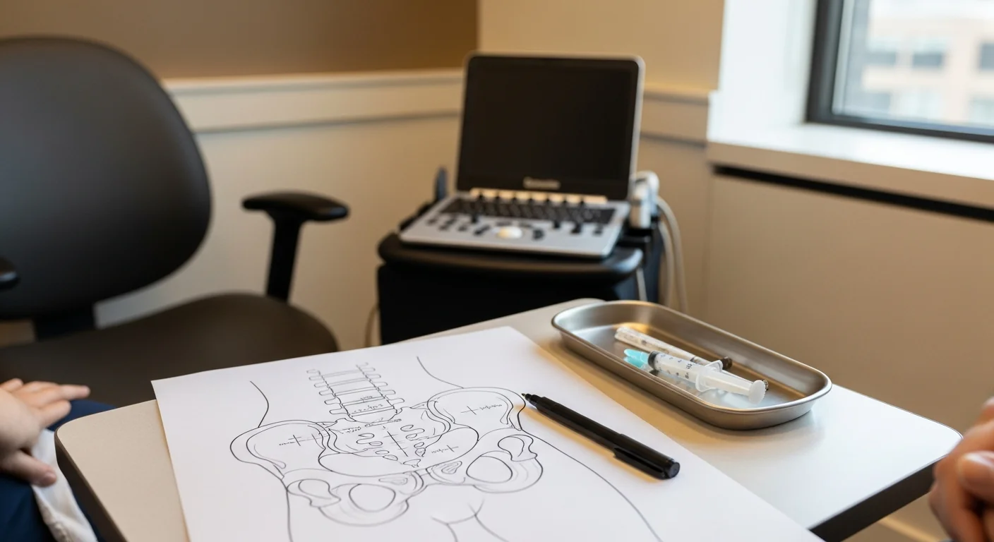
Every diagnostic visit begins with a cutaneous map. The patient marks the painful area on a body diagram with a fine-tip marker. The dermatomal map is the single most useful diagnostic artifact — a band that respects the intercostobrachial territory (axilla to medial elbow) tells you immediately which block to run, and a defined patch confined to the anterolateral thigh tells you LFCN regardless of what the surgical report shows.
The second move is the Tinel test. Gentle percussion along the surgical scar and at the candidate entrapment points reproduces neuropathic pain in a positive case. The Tinel-positive point is the target for the diagnostic block. The third move is provocative testing — Carnett’s sign for abdominal wall pain, FAIR test for piriformis-pattern pain after hip surgery, Spurling for cervical components after thoracic outlet–pattern syndromes.
Diagnostic nerve blocks under ultrasound or fluoroscopy as the gold standard
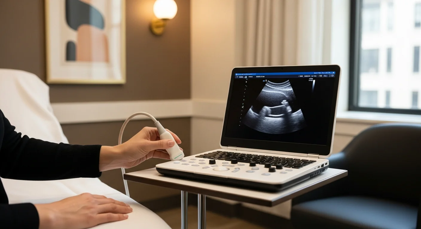
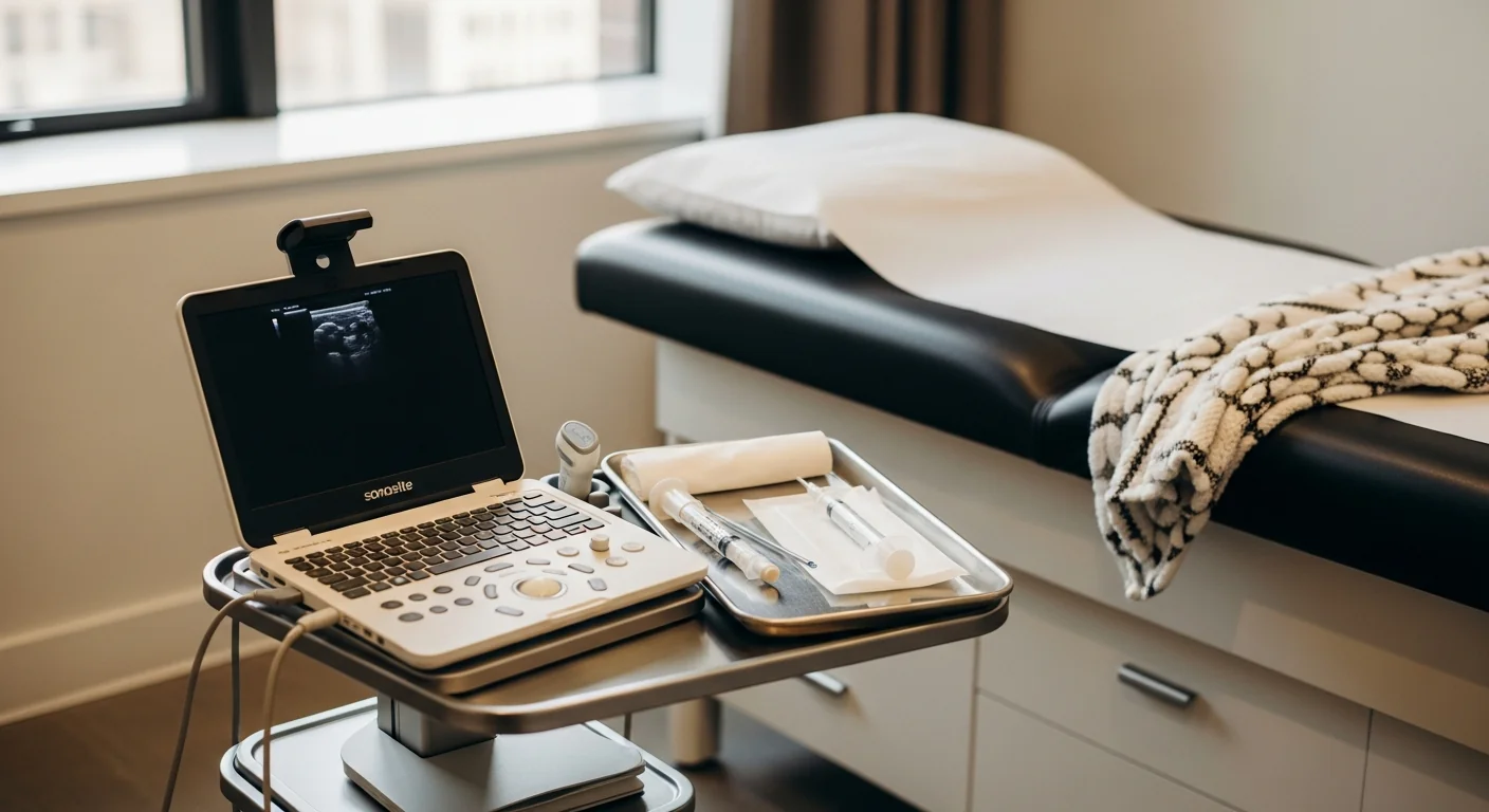
The diagnostic peripheral nerve block is the single single most useful tool in post-surgical pain medicine. Under ultrasound (for superficial peripheral nerves) or fluoroscopy (for axial and deeper paravertebral targets), a small volume of local anesthetic is delivered onto or immediately adjacent to the suspected nerve. The diagnostic answer arrives in 10–15 minutes. ≥50% pain abolition in the working window is a positive block; <50% redirects the workup to a different nerve or a different mechanism.
Image guidance is essential. Blind landmark technique runs accuracy in the 50–70% range for most peripheral nerves; ultrasound and fluoroscopy push accuracy above 95%. In surgically distorted anatomy — which is by definition the case for every iatrogenic neuropathy — blind technique is inadequate and the diagnostic ambiguity it produces propagates into wasted procedural attempts and incorrect surgical referrals.
When advanced imaging adds value (and when it doesn’t)
High-resolution musculoskeletal ultrasound is the first-line imaging tool — performed at the consultation, identifies neuromas and perineural scar, and directly guides the diagnostic block. MR neurography (a dedicated peripheral-nerve MRI protocol) is useful for proximal lesions and for documenting nerve thickening before surgical referral. Standard MRI of the operative site rarely changes treatment unless the question is recurrent disc, mesh malposition, or implant failure — and those are surgical questions, not pain-medicine questions.
The decision tree above (When to consider pain medicine evaluation after surgery) walks through the timing markers we use at Modal Pain to triage post-surgical patients. The 12-week mark is where chronic post-surgical pain becomes the formal diagnosis and where image-guided diagnostic blocks become the highest-impact next step.
Treatment ladder
Image-guided diagnostic blocks (often therapeutic)
The diagnostic block is the workhorse. A small volume of local anesthetic — typically 3–5 mL of bupivacaine — delivered under ultrasound or fluoroscopic guidance onto the suspected nerve. Diagnostic in 10–15 minutes; therapeutic for 4–12 weeks with corticosteroid added. The same procedure is repeatable. See the dedicated nerve blocks page for the full catalog of blocks we perform.
Hydrodissection for perineural fibrosis
For nerves tethered in dense surgical scar, ultrasound-guided hydrodissection mechanically separates the nerve from the surrounding fibrosis using volume injection of saline, 5% dextrose, or dilute corticosteroid. Restores nerve gliding, often produces dramatic and durable relief, particularly in post-hernia, post-Pfannenstiel, and post-mastectomy scar.
Radiofrequency ablation for refractory cases
For patients who respond clearly to diagnostic blocks but get diminishing duration with each repeat, pulsed radiofrequency ablation at the same target produces 6–12 months of relief. Pulsed (not conventional thermal) is essential for most iatrogenic neuropathies — the lesion must be neuromodulatory rather than destructive to avoid creating deafferentation pain in cosmetically or functionally sensitive territory.
Cryoneurolysis for selected intercostal and peripheral targets
Percutaneous cryoablation produces dense sensory blockade lasting 6–12+ months by freezing the nerve, which Wallerian-degenerates and regenerates predictably. Particularly useful for intercostal neuralgia after thoracotomy or rib surgery. Modal Pain coordinates referral for cryoablation when the standard pulsed-RFA ladder reaches its ceiling.
Surgical referral when conservative care fails
For severe refractory cases, surgical neurectomy, nerve decompression, or modern neuroma management (RPNI, TMR) by a peripheral nerve specialist is the durable salvage option. Specific procedures we coordinate referral to:
- Amid triple neurectomy — for refractory post-hernia inguinodynia, performed by peripheral nerve plastic surgeons.
- Anterior neurectomy at the rectus sheath — for ACNES and post-laparoscopic-port abdominal wall neuralgia.
- RPNI (regenerative peripheral nerve interface) — for terminal neuromas after amputation or major peripheral nerve injury; the regenerating axon is implanted into a small denervated muscle graft.
- TMR (targeted muscle reinnervation) — for neuromas at major nerve transection sites; the proximal nerve stump is coapted to a motor nerve of a small expendable muscle.
- Dellon-style nerve decompression — for compressive neuropathy at named entrapment sites (tarsal tunnel, Wartenberg’s, cubital tunnel).
- Microvascular decompression — for cranial neuralgia with confirmed vascular contact (see the facial and cranial nerve pain page).
Modal Pain coordinates these referrals directly when indicated. We provide the surgical team with the documented diagnostic block response and imaging that surgical neurectomy programs require before accepting a referral. Naming individual surgeons publicly is something Modal Pain does not do; the referral network is selected case-by-case based on the patient’s diagnosis, geography, and insurance.
Before considering a second surgery
A meaningful minority of post-surgical pain patients are referred back to their original surgeon for “exploration” or revision surgery before a peripheral nerve workup has been done. The probability that revision in the same surgical field will resolve a chronic post-surgical neuropathic pain is low — and the risk of generating additional iatrogenic nerve injury in the same field is real.
The decision rule above walks through the six-step process we use to triage patients being offered revision surgery. Most often, the right answer is to identify and treat the peripheral nerve generator directly — not to operate again in the original field.
Frequently asked questions
The FAQ block below ships its own schema via the page route; questions are tuned to the actual patient and referring-physician queries we receive at Modal Pain on post-surgical pain.
Related articles
- Chronic Groin Pain After Hernia Surgery: When the Nerve Got Caught
- C-Section Scar Pain That Won’t Go Away: When Nerves Get Trapped in the Pfannenstiel Incision
- Chronic Pelvic Pain After Hysterectomy: When Nerves Get Caught in the Surgical Field
- Post-Mastectomy Nerve Pain: The Intercostobrachial Neuralgia You Were Probably Never Warned About
Ready to evaluate your post-surgical pain
If pain has persisted more than 12 weeks after a surgery and is not improving, an image-guided diagnostic evaluation is the next step. Same-week new-patient consultations are routinely available at 369 Lexington Avenue, Floor 25 in Midtown Manhattan.
Verify your insurance covers a post-surgical nerve pain workup Same-week diagnostic block appointment
Or call (646) 290-6660. Dr. Movshis sees every patient personally — initial consultation through follow-up.