A patient in her late forties has spent two years being treated for a stiff neck. Chiropractic, massage, a cervical MRI showing “age-appropriate degeneration,” a course of physical therapy that helped for a week at a time. Her head drifts to the right when she is tired, her right sternocleidomastoid is thick and sore, and when she rests two fingers against her chin the pull eases — a detail she has never mentioned because it seemed strange. That is cervical dystonia, and it is the most common adult-onset focal dystonia in the world. In one U.S. registry study the average patient waited 43.7 months and saw 3.5 providers before anyone named it (Tiderington 2013).
Dr. Alex Movshis treats cervical dystonia at Modal Pain Management, 369 Lexington Avenue in Midtown Manhattan, with EMG-guided injections of Botox™, Dysport™, or Daxxify™ into the specific muscles that are pulling, adding ultrasound for the deep muscles. This page covers how the condition is recognized, how it is diagnosed, why it gets missed, what the injections involve, what else helps, and what to expect from treatment here.
What cervical dystonia is, and what it is not
Cervical dystonia is a neurological movement disorder. The brain’s motor network — basal ganglia, cerebellum, and the sensorimotor cortex — sends a sustained or intermittent, involuntary contraction command to a set of neck muscles, and the head is pulled into a posture the patient did not choose and cannot fully override. It is not a spine problem, although the years of abnormal loading eventually produce one. It is not a psychological problem, although a century ago it was labeled one. It is not a muscle strain, although the strained-muscle treatments people receive for it do briefly soothe the secondary ache.
Onset is usually between 40 and 60. Women are affected about one and a half times as often as men. Most cases are idiopathic — no cause is ever found — and a family history of dystonia or tremor is present in a minority. A small number are secondary: dopamine-blocking drugs (metoclopramide, prochlorperazine, most antipsychotics) can produce an identical picture called tardive dystonia, Wilson disease has to be excluded in anyone with onset under 40, and a structural lesion of the upper cervical spine or brainstem is a rare cause that imaging identifies.
Three features distinguish it from every other cause of neck pain we see. The first is the sensory trick, or geste antagoniste: a light touch to the chin, cheek, or back of the head that reduces the pull far more than the mechanical force of the touch could explain. Most patients have one and few volunteer it. The second is a head tremor that is jerky, irregular, and worse when the patient tries to hold the head straight against the pull — different from the smooth, symmetrical tremor of essential tremor, and often the reason a patient is told they have “a tremor” rather than dystonia. The third is pain that sits in the muscles doing the pulling and improves when those muscles are quieted, which is the reverse of most degenerative neck pain.
The four head positions and why the muscle map matters
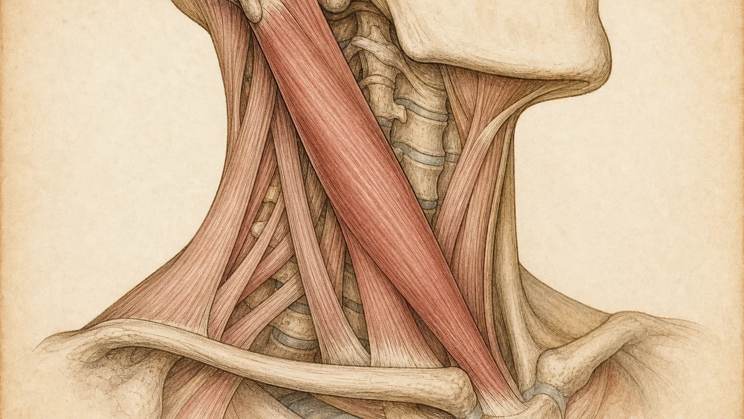
The direction of the pull tells you which muscles are firing, and that determines the injection. Rotation of the chin toward one shoulder is torticollis, the most common pattern, driven mainly by the sternocleidomastoid on the side opposite the chin and the splenius capitis on the same side as the chin. A tilt of the ear toward the shoulder is laterocollis, driven by the sternocleidomastoid, splenius, levator scapulae, and scalenes on the side the ear drops toward. A forward pull of the chin toward the chest is anterocollis, driven by both sternocleidomastoids, the scalenes, and the deep longus colli, and it is the hardest pattern to treat well. A backward pull is retrocollis, driven by the splenius and semispinalis on both sides and the upper trapezius. Sideways and forward shifts of the whole head add two more patterns, and most patients have a combination.
The 2013 consensus classification of dystonia (Albanese 2013) and the “collis versus caput” distinction refined the map further: a rotation that happens at the skull-to-upper-neck level (caput) implicates different muscles — the obliquus capitis inferior in particular — than a rotation that happens lower in the neck (collis). Injecting the wrong level is the commonest reason a competent-looking injection does nothing. This is why the examination is repeated before every cycle, not just the first, and why guidance with EMG and ultrasound is not optional for the deep muscles.
How cervical dystonia is diagnosed
The diagnosis is made in the exam room, not the scanner. Dr. Movshis watches the head at rest, during conversation, while walking, and while the patient tries to hold it straight, then asks the patient to use their trick and watches what changes. The neck muscles are palpated one by one for tone, tenderness, and hypertrophy. Range of motion is measured in each plane against the pull and with it. Tremor is characterized by direction and regularity. The rest of the body is examined for dystonia elsewhere (eyelids, jaw, hand) and for parkinsonism, because either changes the diagnosis and the referral. The severity of the posture, the disability, and the pain are scored on the Toronto Western Spasmodic Torticollis Rating Scale (TWSTRS), which is the scale used in every trial cited on this page and the one we use to measure your response cycle over cycle.
Tests are ordered to rule things out, not in. A cervical spine MRI is appropriate when there is arm pain, numbness, or weakness that suggests a pinched nerve in the neck, or when the posture could be explained by a structural problem. A brain MRI is appropriate when onset is young, the dystonia is spreading, or the exam shows anything beyond the neck. A serum ceruloplasmin is drawn for anyone under 40. Blood tests are otherwise unremarkable and EMG at the diagnostic stage is used to characterize the pattern, not to confirm the disease.
Why it gets missed for years
Every patient with cervical dystonia has neck pain, and neck pain has a well-worn pathway: anti-inflammatories, a chiropractor, massage, physical therapy, an MRI that finds the degeneration present in most adults over 40, and a diagnosis of cervical spondylosis or muscle strain. Each step helps a little because the secondary muscle pain responds to anything that relaxes muscle. The improvement wears off, the head keeps drifting, and the patient is told to stretch more. The sensory trick, which would settle the question in thirty seconds, goes unmentioned because patients assume it is odd, and unasked because most clinicians have never been taught to ask.
The result is the 43.7-month delay documented by Tiderington and colleagues, during which 63% of patients tried oral medications that mostly did not work, and the muscles kept shortening. Long-standing untreated dystonia produces real secondary damage: accelerated facet arthritis on the side of rotation, disc degeneration at the levels doing the most abnormal work, and in some cases a cervical radiculopathy. Those problems are treatable — see the neck pain page for how — but they are avoidable if the dystonia is treated when it starts. If you have already recognized the early pattern, our guide to the early symptoms of cervical dystonia walks through what to watch for and when to be seen.
Botulinum toxin: the first-line treatment, and how it is done here
Botulinum toxin blocks the release of acetylcholine at the junction between nerve and muscle. Injected in small volumes into an overactive muscle, it weakens that muscle for roughly three months without affecting the muscles around it. In cervical dystonia that is exactly the mechanism needed: quiet the muscles that are pulling, leave the antagonists alone, and let the head return toward midline. In the 2020 Cochrane review of nine randomized trials (1,144 participants), a single session improved the TWSTRS total score by 8.09 points versus placebo at week 4, about 18% from baseline, and the pain subscore by 2.11 points, with no efficacy difference between the type-A products (Rodrigues 2020). The 2016 American Academy of Neurology guideline, since retired by the AAN, had rated abobotulinumtoxinA (Dysport™) and rimabotulinumtoxinB (Myobloc™) as established effective and onabotulinumtoxinA (Botox™) and incobotulinumtoxinA (Xeomin™) as probably effective — a difference that reflected trial design, not clinical performance (Simpson 2016). DaxibotulinumtoxinA (Daxxify™) was approved for cervical dystonia in 2023 on the ASPEN-1 trial, in which the 125-unit dose improved the TWSTRS total score by 12.7 points versus 4.3 for placebo at weeks 4–6, with a median duration of effect of 24 weeks (Comella 2024).
Prefer to watch? A 95-second narrated animation of the mechanism — the order without a brake, the touch that eases it, and what botulinum toxin does at the nerve ending — with the full transcript and PubMed-verified references.
At Modal Pain Management the procedure is built around the muscle map. Dr. Movshis re-examines the pattern at the start of every visit, selects the muscles and the dose for each, and injects under EMG guidance every time, which confirms the needle is inside a muscle that is actively firing, adding ultrasound when the map includes deep muscles, so the needle reaches them — the longus colli, the scalenes, the levator scapulae, and the obliquus capitis inferior — without passing through the carotid sheath or the brachial plexus. The choice between Botox™, Dysport™, and Daxxify™ depends on your pattern, your response to prior product, and how long you need each cycle to last. Dysport’s units are not interchangeable with Botox units. Daxxify’s longer duration suits patients whose benefit fades before the twelfth week.
The effect begins within the first two weeks, peaks at four to six weeks, and wears off at 10–14 weeks with Botox™ or Dysport™, so cycles are scheduled at 12 weeks. What the visit involves, how the units are decided, what to expect after the injections, and what it costs are covered in Botox for cervical dystonia: what to expect. The side effect that matters is difficulty swallowing, which follows toxin spreading from the sternocleidomastoid to the swallowing muscles. The Botox™ prescribing information reports it in 19% of cervical dystonia patients and warns it can last for months. In a long-term clinic series it was usually mild and short-lasting (Yahalom 2021). It is why the dose into the sternocleidomastoid, especially both sides at once, is kept deliberate. Neck weakness, a flu-like day after the first session, and injection-site soreness are the other common effects. In ASPEN-1, dysphagia occurred in 1.6% of patients at the 125-unit Daxxify dose (Comella 2024).
When the injections do not work
Roughly one patient in four gets less benefit than expected from a given cycle, and the reason is almost always mechanical rather than biological. The pattern has shifted since the last map. A deep muscle is contributing and has never been injected. The dose in the main mover is too low, or the injection landed in the wrong level of a two-level rotation. Anterocollis often fails when the longus colli is left alone, and we reach it under ultrasound, which shows the carotid sheath and the nerves beside it. Neutralizing antibodies, which made toxin stop working in a meaningful fraction of patients treated with the original high-protein formulations in the 1990s, are rare with current products.
The response is a re-map under EMG and ultrasound, a dose adjustment, and if needed a switch of product. Patients who do not respond to two well-executed products over two cycles each are candidates for bilateral deep brain stimulation of the globus pallidus internus. In the only sham-controlled trial, pallidal stimulation improved the TWSTRS severity score by 5.1 points at three months against 1.3 points with sham stimulation, in patients who had failed medication and toxin (Volkmann 2014). Selective peripheral denervation, the older surgical option, is now rarely performed. We coordinate that referral rather than continuing injections that are not working.
What else helps: therapy, medication, and treating the secondary pain
Physical therapy changes the arithmetic of the injections. In a randomized trial, patients who received botulinum toxin plus a structured neuromotor rehabilitation program improved more and needed lower toxin doses over successive cycles than patients who received toxin alone (Tassorelli 2006). The program is not generic neck stretching. It is built around the pattern — active correction toward midline using the sensory trick as a cue, strengthening of the antagonist muscles, posture and breathing work — and it starts in the second or third week after each injection, when the treated muscles are quiet enough to retrain. The program runs with Modal’s in-house physical therapists, on the same floor, so it can be timed to each injection cycle. See our physical therapy page for how it works.
Oral medications have a smaller role than patients expect. Trihexyphenidyl, clonazepam, and baclofen each help a minority of patients, and each is sedating or causes dry mouth, constipation, or memory complaints at the doses that help. They are used as a bridge while an authorization is pending, as an adjunct for tremor, or in the few patients who cannot receive toxin.
The secondary pain has its own treatments. Years of one-sided pull produce myofascial trigger points in the trapezius and levator scapulae that respond to trigger point injections, facet joint arthritis on the side of rotation that responds to a medial branch block and, when the block confirms the source, radiofrequency ablation, and occasionally a cervical radiculopathy that responds to a cervical epidural steroid injection. Headache that starts at the base of the skull on the side of the pull is often cervicogenic rather than migraine — our cervicogenic headache film shows the mechanism — and an occipital nerve block both tests and treats it. In registry data from more than a thousand patients, the pain component tracked disability more closely than the posture did (Charles 2014), which is why it is treated alongside the dystonia rather than after it.
Daily life: sleep, driving, work, and the questions patients ask
The pull is worst when tired, stressed, or walking, and eases lying down, which is why most patients sleep well and struggle in the afternoon. Driving is usually possible once the injections hold the head near midline, and a head rest set to the correct height helps between cycles. Desk work is manageable with the monitor placed slightly toward the side the head prefers, so the dystonic pull works with the task rather than against it. Sensory tricks can be engineered: a collar, a scarf, or a pair of glasses with a wide temple that touches the cheek reproduces the effect of a hand for some patients.
Cervical dystonia does not shorten life. It can qualify as a disability when the documented functional limitation meets the relevant criteria, which depends on the individual case rather than the diagnosis alone, and we provide the clinical documentation when a patient applies. Anxiety and low mood are common alongside it — partly reactive, partly part of the disorder — and are worth naming at the visit, because treating them improves the response to everything else.
What to expect at Modal Pain Management
The first visit is a consultation that takes 45–60 minutes and may include an ultrasound exam. Bring any prior imaging and the list of everything you have tried, including any dopamine-blocking medication in your history. Dr. Movshis examines the pattern, scores it, explains which muscles are involved, and, if the diagnosis is cervical dystonia, recommends a product and a map. Prior authorization is submitted the same day, typically takes 7–14 business days, and the first injection is generally scheduled within 2–3 weeks. The injection visit takes 20–30 minutes. You drive yourself home and can go back to work the same day. For the first 24 hours, skip workouts, neck massage, heat on the neck and over-the-counter anti-inflammatories. A follow-up about two weeks later records the response and any side effect on the same scale, and the map is adjusted before the next cycle. Botox™, Dysport™, and Daxxify™ for cervical dystonia are covered by most commercial PPO plans with prior authorization. Modal Pain Management does not accept Medicare, Medicaid, HMO plans, or workers’ compensation. Self-pay pricing for each product is published per unit, and you can verify your insurance before the visit.
If your head does not stay where you put it, that is the visit to book.
References
- Rodrigues FB, Duarte GS, Marques RE, et al. Botulinum toxin type A therapy for cervical dystonia. Cochrane Database Syst Rev. 2020;11(11):CD003633. doi:10.1002/14651858.CD003633.pub4
- Simpson DM, Hallett M, Ashman EJ, et al. Practice guideline update summary: Botulinum neurotoxin for the treatment of blepharospasm, cervical dystonia, adult spasticity, and headache [retired by the AAN]. Neurology. 2016;86(19):1818–1826. doi:10.1212/WNL.0000000000002560
- Tiderington E, Goodman EM, Rosen AR, et al. How long does it take to diagnose cervical dystonia? J Neurol Sci. 2013;335(1–2):72–74. doi:10.1016/j.jns.2013.08.028
- Albanese A, Bhatia K, Bressman SB, et al. Phenomenology and classification of dystonia: a consensus update. Mov Disord. 2013;28(7):863–873. doi:10.1002/mds.25475
- Comella CL, Jankovic J, Hauser RA, et al. Efficacy and safety of daxibotulinumtoxinA for injection in cervical dystonia: ASPEN-1 phase 3 randomized controlled trial. Neurology. 2024;102(4):e208091. doi:10.1212/WNL.0000000000208091
- Tassorelli C, Mancini F, Balloni L, et al. Botulinum toxin and neuromotor rehabilitation: an integrated approach to idiopathic cervical dystonia. Mov Disord. 2006;21(12):2240–2243. doi:10.1002/mds.21145
- Volkmann J, Mueller J, Deuschl G, et al. Pallidal neurostimulation in patients with medication-refractory cervical dystonia: a randomised, sham-controlled trial. Lancet Neurol. 2014;13(9):875–884. doi:10.1016/S1474-4422(14)70143-7
- Charles PD, Adler CH, Stacy M, et al. Cervical dystonia and pain: characteristics and treatment patterns from CD PROBE. J Neurol. 2014;261(7):1309–1319. doi:10.1007/s00415-014-7343-6
- Yahalom G, Fay-Karmon T, Livneh V, et al. Botulinum injections for idiopathic cervical dystonia: a longitudinal study. Neurotox Res. 2021;39(4):1352–1359. doi:10.1007/s12640-021-00378-2 · PubMed