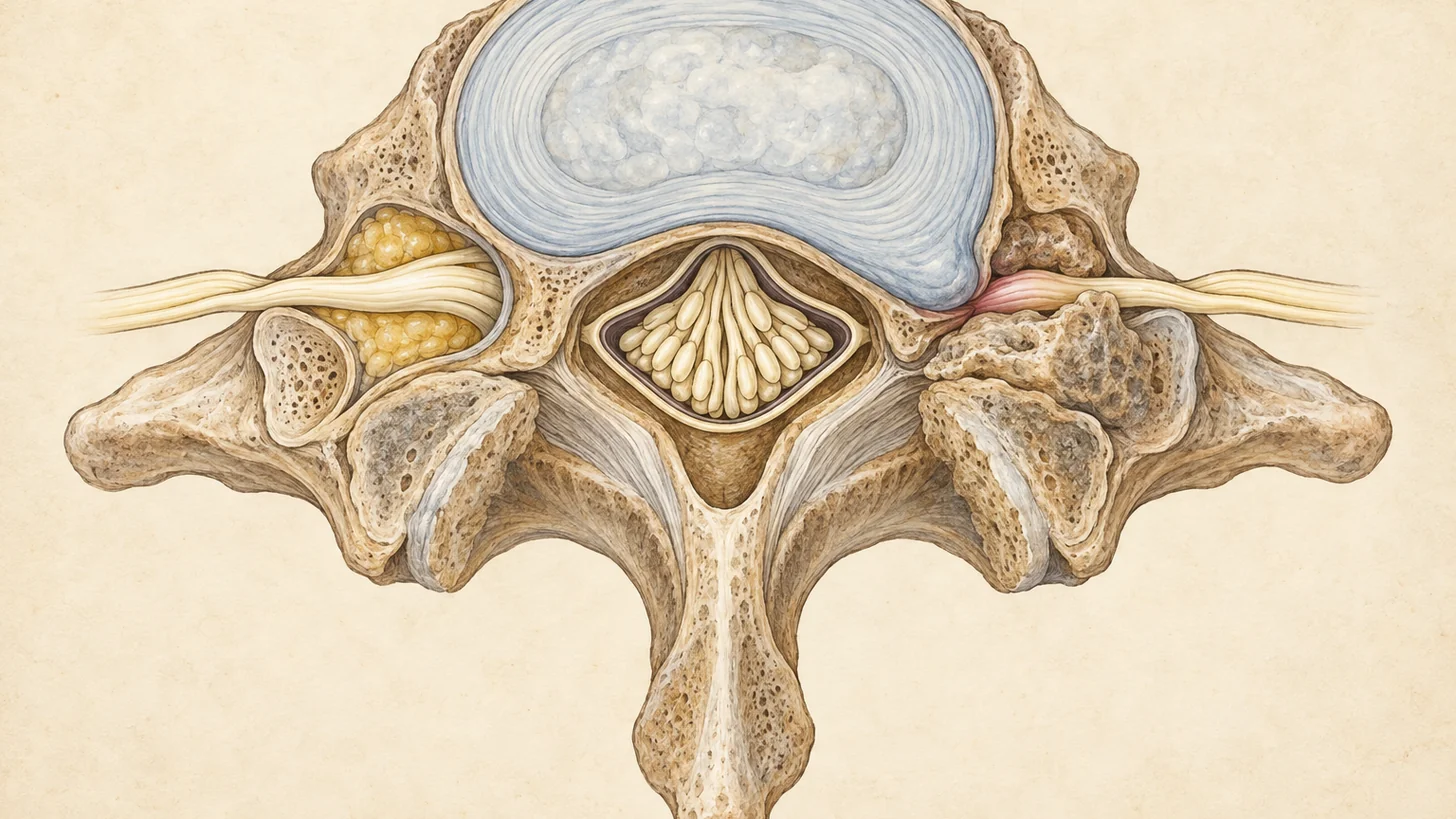
Radiculopathy — the clinical term for a “pinched nerve” at the spine — occurs when a nerve root is compressed or inflamed as it exits the spinal column, producing pain, numbness, tingling, or weakness that travels into the arm or leg along a predictable nerve pathway. At Modal Pain Management, at 369 Lexington Avenue, Floor 25 in Midtown Manhattan (one block from Grand Central Terminal), Dr. Alex Movshis, MD — dual board-certified in anesthesiology and pain medicine, fellowship-trained in interventional pain medicine at the Icahn School of Medicine at Mount Sinai — diagnoses and treats cervical and lumbar radiculopathy with image-guided, non-opioid interventional care. Same-week appointments are available. Call (646) 290-6660 or book a consultation online.
Prefer to watch? An 88-second 3D animation of a pinched nerve in the neck — the root’s bony doorway, the finger map, why looking up makes it worse, and the signs that the spinal cord is involved — with the full transcript and PubMed-verified references.
What Is Radiculopathy? Cervical, Thoracic, and Lumbar
Radiculopathy is common — population data put the annual incidence of cervical radiculopathy alone at roughly 83 per 100,000 people, peaking between ages 50 and 54 (Radhakrishnan et al., Brain, 1994). The good news: most cases improve without surgery. In that same population study, 90% of patients were symptom-free or only mildly limited at follow-up, though about a third had a recurrence and 26% eventually had surgery (Radhakrishnan 1994).
Every spinal nerve root exits the spinal canal through a bony opening called the neural foramen. When that foramen narrows, or when disc material, bone spurs, or inflamed tissue encroach on the root, the nerve misfires. Because each root supplies a defined strip of skin (a dermatome) and a defined group of muscles (a myotome), radiculopathy produces a recognizable pattern — not vague “pain everywhere,” but symptoms in a specific distribution. That pattern is what lets a trained examiner localize the problem, often before any imaging is ordered (StatPearls: Radiculopathy, NCBI Bookshelf).
Cervical radiculopathy affects the nerve roots of the neck (C5–C8 most commonly) and sends symptoms into the shoulder, arm, forearm, hand, or specific fingers — often a deep ache at the shoulder blade plus electric or burning pain down the arm, sometimes with grip weakness. It frequently coexists with neck pain, though the arm symptoms usually dominate. C7 is the root most often involved, followed by C6 (Radhakrishnan 1994).
Lumbar radiculopathy affects the nerve roots of the lower back (L4, L5, and S1 most commonly) and sends symptoms into the buttock, thigh, calf, or foot. Radicular pain traveling down the leg in an L4–S1 pattern is what most people call sciatica, which is why our sciatica page and this page overlap: sciatica is best understood as a symptom of lumbar radiculopathy, not a separate diagnosis. Low back pain with leg pain below the knee affects an estimated 9.9% to 25% of the general population in a given year (Van Boxem 2010). Leg pain typically exceeds back pain when a nerve root is the true generator.
Thoracic radiculopathy is uncommon and produces a band-like pattern of pain or numbness wrapping around the chest or upper abdomen. Because it can mimic cardiac, gastrointestinal, or shingles pain, it warrants careful evaluation before being attributed to the spine.
Radiculopathy at a nerve root is also distinct from peripheral nerve entrapment — compression farther out in the limb, such as carpal tunnel syndrome. The two can coexist (a “double crush”), and telling them apart is one reason electrodiagnostic testing matters. Modal Pain also evaluates peripheral nerve entrapment when the exam points away from the spine.
Common Causes of a Pinched Nerve
Most radiculopathy comes from a short list of structural problems, and the likely cause shifts with age: acute disc herniation is more common in younger adults, and degenerative foraminal narrowing in older ones (StatPearls: Radiculopathy, NCBI Bookshelf).
- Herniated disc — the disc’s inner nucleus pushes through its outer layer and compresses or chemically inflames an adjacent root. It is the most common cause of acute radiculopathy and has a good natural history. Extrusions and sequestrations shrink on follow-up MRI in most cases, while contained protrusions shrink less often (Chiu 2015).
- Foraminal stenosis — disc height loss, facet arthritis, and bone spurs narrow the neural foramen and crowd the exiting root (what the grades on an MRI report mean is explained in our guide to foraminal stenosis on MRI). Symptoms build gradually and worsen with standing or spinal extension.
- Degenerative disc disease and spondylosis — chronic arthritic change can irritate a root even without a discrete herniation. In the neck, spondylosis or disc disease accounted for 68% of cases in the population study above, and a discrete disc protrusion for 22% (Radhakrishnan 1994).
- Spondylolisthesis — one vertebra slips forward on the next, kinking the foramen and tethering the root.
- Central spinal stenosis — canal narrowing compresses multiple roots at once, classically causing leg heaviness with walking that eases with sitting or leaning forward (neurogenic claudication). In the neck, the same narrowing can press on the spinal cord as well as the roots, which is covered on the cervical stenosis page.
- Less common causes — post-surgical scar tissue (see post-laminectomy syndrome), diabetic radiculoplexopathy, infection, and tumor. These are why “it’s just a pinched nerve” should be a diagnosis made after evaluation, not before.
Symptoms by Nerve Root: How the Pattern Localizes the Problem
The distribution of symptoms is the single most useful clinical clue. The table below summarizes the classic presentation of the most commonly affected roots, consistent with standard dermatomal references (Cleveland Clinic: Radiculopathy). Individual anatomy varies — the exam, not the table, makes the diagnosis.
| Nerve Root | Pain / Numbness Distribution | Typical Weakness | Reflex Change |
|---|---|---|---|
| C5 | Neck, shoulder, lateral upper arm | Shoulder abduction, elbow flexion | Biceps |
| C6 | Lateral forearm, thumb, index finger | Wrist extension, elbow flexion | Brachioradialis |
| C7 | Posterior arm, middle finger | Elbow extension, wrist flexion | Triceps |
| C8 | Medial forearm, ring and little fingers | Finger flexion, grip | None reliably |
| L4 | Anterior thigh, inner knee, medial shin | Knee extension, hip flexion | Patellar |
| L5 | Lateral thigh and calf, top of foot, big toe | Foot and big-toe dorsiflexion (foot drop in severe cases) | None reliably |
| S1 | Back of thigh and calf, outer foot, little toe | Foot plantarflexion, toe walking | Achilles |
Red-Flag Symptoms That Need Urgent Care
Most radiculopathy is painful but not dangerous. A small number of presentations are emergencies, and we screen for them at every visit:
- New bowel or bladder dysfunction — urinary retention, overflow incontinence, or loss of rectal tone, especially with numbness in the saddle region. This suggests cauda equina syndrome and requires same-day emergency evaluation.
- Progressive or profound weakness — a rapidly worsening foot drop or grip failure is a surgical urgency, not a wait-and-see problem.
- Bilateral leg symptoms with gait instability or hand clumsiness — may indicate spinal cord compression (myelopathy) rather than a single pinched root.
- Fever, unexplained weight loss, night pain unrelieved by position change, recent cancer, or immunosuppression — raises concern for infection or malignancy and changes the workup entirely.
If any of these apply to you, do not wait for a routine appointment: call 911 or go to the nearest emergency room. Our office can help with follow-up afterward at (646) 290-6660.
Diagnosis and Workup at Modal Pain Management
Accurate treatment starts with confirming which root is involved, what is compressing it, and whether the nerve is actually the pain generator. Dr. Movshis approaches the workup in stages.
History and Neurological Examination
Your visit begins with a detailed history — onset, mechanism, positions that provoke or relieve symptoms, prior episodes and treatments — followed by a structured neurological examination: muscle strength graded against resistance, reflexes, sensory mapping, and gait. Provocative maneuvers help reproduce the pattern. The straight-leg-raise test tensions the L4–S1 roots and can reproduce radiating leg pain below the knee. Spurling’s maneuver, gentle neck extension and rotation toward the symptomatic side, narrows the cervical foramen and can reproduce arm symptoms. A positive provocative test in a matching dermatomal pattern substantially raises diagnostic confidence before any imaging is ordered (StatPearls: Radiculopathy, NCBI Bookshelf).
Imaging: MRI First, Matched to the Exam
MRI without contrast is the preferred initial study for suspected radiculopathy because it shows disc, nerve root, and soft-tissue anatomy directly. Current appropriateness criteria support MRI when radicular symptoms persist beyond roughly six weeks of conservative care, when progressive neurological deficit is present, or when an intervention or surgery is being planned (ACR Appropriateness Criteria: Low Back Pain, American College of Radiology). Just as important as ordering the scan is reading it against your exam. Disc bulges appear in 30% of pain-free 20-year-olds and 84% of pain-free 80-year-olds (Brinjikji 2015), so a “positive” MRI only matters if the compressed level matches your dermatomal pattern. CT is reserved for patients who cannot undergo MRI. X-rays assess alignment but cannot show nerve roots.
Electrodiagnostic Testing (EMG/NCS)
Nerve conduction studies and electromyography are used selectively — when the picture is ambiguous: to distinguish radiculopathy from peripheral entrapment (carpal tunnel, peroneal neuropathy), to confirm double-crush overlap, to grade severity, or to document the diagnosis when imaging and exam disagree. EMG can detect denervation changes MRI cannot, but it has limited sensitivity for pain-only radiculopathy, so a normal EMG does not rule the diagnosis out.
Diagnostic Selective Nerve Root Block
When imaging shows multilevel degeneration and it is unclear which root is generating your symptoms — a common situation, since most MRI reports list findings at several levels — a diagnostic selective nerve root block can help. Under fluoroscopic guidance, a small volume of local anesthetic is placed around one suspected root. If your familiar pain drops sharply during the anesthetic window, that root becomes the likely source. If nothing changes, attention shifts elsewhere. The point is to avoid treating the level that looks worst on MRI rather than the level that actually hurts. The 2010 evidence-based guideline on lumbosacral radicular pain describes selective blocks as helping to confirm the affected level (Van Boxem 2010).
Treatment Options for Radiculopathy
Treatment is staged: conservative care first for most patients, interventional procedures when conservative care is not enough, and surgical referral when the neurological findings demand it. Our non-opioid philosophy means your plan treats the inflamed nerve root directly rather than masking it with systemic medication.
Conservative Care and Physical Therapy
Because most acute radiculopathy improves substantially over 4–12 weeks as herniations resorb and inflammation settles, the first phase is usually activity modification, a short course of anti-inflammatory or neuropathic medication where appropriate, and structured physical therapy — directional-preference exercises, nerve-glide techniques, and progressive core or scapular stabilization. Staying active within tolerance is favored over bed rest, which delays recovery (StatPearls: Radiculopathy, NCBI Bookshelf). Modal’s in-house physical therapists work on the same floor, and chiropractic coordination is available under the same roof at our Lexington Avenue office.
Epidural Steroid Injections
When radicular pain persists beyond several weeks of conservative care, an epidural steroid injection (ESI) delivers anti-inflammatory corticosteroid directly to the epidural space around the irritated nerve root under real-time fluoroscopic guidance. Dr. Movshis selects the approach — interlaminar, transforaminal, or caudal — based on your imaging and symptom distribution. A large systematic review and meta-analysis found that ESIs provide a small short-term improvement in radicular leg pain and function, below what the authors set as clinically important, with no longer-term benefit (Chou 2015). A blinded trial of transforaminal injections gave a more favorable picture: 54% of patients given steroid had at least half their leg pain relieved at one month, against 7% to 21% in control groups, though fewer kept that relief over the following year (Ghahreman 2010). For radicular pain lasting three months or longer, a 2025 BMJ guideline panel recommended against epidural injections (Busse 2025). Injections are best understood as a tool to calm the nerve and enable rehabilitation, not a permanent fix. The epidural steroid injection page linked above covers candidacy criteria, the FDA’s 2014 safety communication on rare neurologic risks (the reviewers’ assessment: Racoosin 2015), and why cervical injections at Modal Pain are interlaminar, with non-particulate steroid.
Selective Nerve Root Blocks
A selective nerve root block places a small volume of anesthetic (with or without steroid) precisely at one exiting root. It serves two roles: diagnostic, helping to show which level is the likely pain source when MRI and exam are ambiguous, and therapeutic, delivering medication to a single symptomatic root, which can be useful for foraminal stenosis. The 2010 evidence-based guideline on lumbosacral radicular pain says selective diagnostic blocks help confirm the affected level, and it recommends transforaminal steroid for subacute radicular pain from a contained herniation when conservative care fails (Van Boxem 2010). More on our approach on the nerve blocks page.
When Surgery Is the Right Answer
We do not perform surgery, and most of our patients never need it. But honest candidacy counseling is part of interventional practice. Surgical consultation is appropriate for progressive motor weakness, cauda equina syndrome, cervical myelopathy, or radicular pain that remains disabling after a well-conducted course of conservative and interventional care. In the randomized SPORT trial of lumbar disc herniation, both surgical and non-surgical groups improved substantially over two years, and the differences between them were small and not statistically significant, with heavy crossover limiting firm conclusions (Weinstein 2006). In the Dutch sciatica trial, early surgery relieved leg pain faster, but disability over the first year was similar and 95% of both groups reported recovery at one year (Peul 2007). Careful non-surgical management is a legitimate path for patients without progressive deficit. When referral is indicated, we coordinate with spine surgeons and continue managing your non-surgical care.
What to Expect During Your Visit
Your first appointment begins with a full consultation. Dr. Movshis will review your symptom history and any prior imaging or EMG reports — bring them if you have them, including a printed copy of the report, or email your imaging to info@modalpain.com ahead of your visit — and perform the neurological examination and provocative testing described above. You will leave with a working diagnosis stated in plain language: which nerve root we believe is involved, and why.
If imaging is needed, we will tell you exactly which study and why, and you can complete it at a conveniently located Manhattan imaging center. If an interventional procedure is recommended, we will explain the target, the evidence, the alternatives, and the risks before scheduling. Procedures are performed in our office-based suite under fluoroscopic guidance with sterile technique: the skin is numbed with local anesthetic, the needle is advanced under live X-ray with contrast confirmation, and the medication is deposited at the target. Most procedures take 15–30 minutes, followed by a short monitored recovery period. Because even local anesthesia can transiently affect sensation and balance, we ask that you arrange a ride home after any injection procedure.
Cost and Insurance for Radiculopathy Treatment
Modal Pain Management accepts most commercial PPO plans, including Aetna, Anthem Blue Cross Blue Shield, Cigna, Empire Blue Cross Blue Shield, the Empire Plan, Oscar, Oxford, UMR and UnitedHealthcare. We verify your benefits before your first visit, at no cost or obligation, so you know your expected out-of-pocket responsibility in advance. Your cost depends on your plan’s deductible, coinsurance, and copay structure. Please note that we do not accept Medicare, Medicaid, HMO plans, or workers’ compensation. Self-pay pricing is available for patients without qualifying coverage, and financing options exist for treatment plans. Details are on our insurance and billing page and our self-pay pricing page, or start with a free benefits check.
Recovery and Long-Term Outlook
Recovery from an interventional procedure is typically quick: most patients take it easy the day of the injection, without staying in bed, resume desk work the next day, and avoid strenuous exertion and heavy lifting for 48 hours. The local anesthetic may give hours of relief that then wears off. The steroid works over days, and the result is judged two to four weeks later. Relief duration varies widely. In recent disc herniations, the injection may calm the nerve while the herniation settles on its own. Full instructions are on the epidural aftercare page.
The long-term outlook for radiculopathy is genuinely favorable for most people. Most acute episodes resolve or become manageable, though recurrences happen, in about a third of people with cervical radiculopathy over five years (Radhakrishnan 1994). Regular exercise, weight management and not smoking are sensible ways to lower the odds. Our role is to calm the nerve root, test the diagnosis when the picture is unclear, and build a plan — with physical therapy and, when appropriate, repeat image-guided treatment — that keeps you functional without opioids and without surgery wherever that is safely possible.
References
- Radhakrishnan K, Litchy WJ, O’Fallon WM, Kurland LT. Epidemiology of cervical radiculopathy. A population-based study from Rochester, Minnesota, 1976 through 1990. Brain. 1994;117 ( Pt 2):325–35. doi:10.1093/brain/117.2.325 · PubMed
- Brinjikji W, Luetmer PH, Comstock B, et al. Systematic literature review of imaging features of spinal degeneration in asymptomatic populations. AJNR Am J Neuroradiol. 2015;36(4):811–6. doi:10.3174/ajnr.A4173 · PubMed
- Van Boxem K, Cheng J, Patijn J, et al. 11. Lumbosacral radicular pain. Pain Pract. 2010;10(4):339–58. doi:10.1111/j.1533-2500.2010.00370.x · PubMed
- Chou R, Hashimoto R, Friedly J, et al. Epidural Corticosteroid Injections for Radiculopathy and Spinal Stenosis: A Systematic Review and Meta-analysis. Ann Intern Med. 2015;163(5):373–81. doi:10.7326/M15-0934 · PubMed
- Ghahreman A, Ferch R, Bogduk N. The efficacy of transforaminal injection of steroids for the treatment of lumbar radicular pain. Pain Med. 2010;11(8):1149–68. doi:10.1111/j.1526-4637.2010.00908.x · PubMed
- Racoosin JA, Seymour SM, Cascio L, Gill R. Serious Neurologic Events after Epidural Glucocorticoid Injection — The FDA’s Risk Assessment. N Engl J Med. 2015;373(24):2299–301. doi:10.1056/NEJMp1511754 · PubMed
- Weinstein JN, Tosteson TD, Lurie JD, et al. Surgical vs nonoperative treatment for lumbar disk herniation: the Spine Patient Outcomes Research Trial (SPORT): a randomized trial. JAMA. 2006;296(20):2441–50. doi:10.1001/jama.296.20.2441 · PubMed
- Peul WC, van Houwelingen HC, van den Hout WB, et al. Surgery versus prolonged conservative treatment for sciatica. N Engl J Med. 2007;356(22):2245–56. doi:10.1056/NEJMoa064039 · PubMed
- Chiu CC, Chuang TY, Chang KH, Wu CH, Lin PW, Hsu WY. The probability of spontaneous regression of lumbar herniated disc: a systematic review. Clin Rehabil. 2015;29(2):184–95. doi:10.1177/0269215514540919 · PubMed
- Busse JW, Genevay S, Agarwal A, et al. Commonly used interventional procedures for non-cancer chronic spine pain: a clinical practice guideline. BMJ. 2025;388:e079970. doi:10.1136/bmj-2024-079970 · PubMed