A herniated disc — also called a slipped or ruptured disc — is one of the most common causes of back pain, neck pain, and radiating arm or leg pain, and the large majority improve without surgery. At Modal Pain Management, at 369 Lexington Avenue, Floor 25, in Midtown Manhattan near Grand Central Terminal, Dr. Alex Movshis — dual board-certified in anesthesiology and pain medicine, Mount Sinai fellowship-trained — specializes in non-surgical, image-guided herniated disc treatment: epidural steroid injections and nerve blocks, coordinated with physical therapy — without opioid medications. Same-week appointments are available, and we verify your commercial PPO benefits before your visit at no charge.
Prefer to watch? A 71-second narrated animation of what a herniated disc actually is — a leak, not a break, and why most shrink on their own — with the full transcript and PubMed-verified references.
What Is a Herniated Disc?
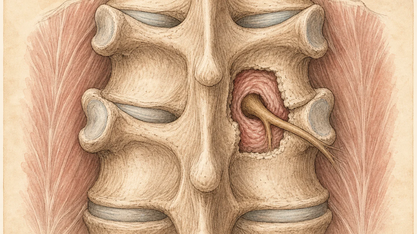
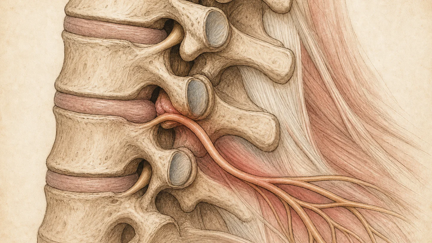
Between each vertebra sits an intervertebral disc — a tough, fibrous outer ring (the annulus fibrosus) surrounding a soft, gel-like center (the nucleus pulposus). A disc “herniates” when the nucleus pushes through a weakened or torn area of the annulus. If the displaced material presses on or chemically irritates a nearby nerve root, it produces the pain, numbness, and weakness that bring patients to our office (StatPearls — Disk Herniation, NCBI).
Clinicians grade disc displacement by how far the material has migrated:
- Disc bulge — the disc extends broadly beyond its boundary, but the outer ring is intact. Bulges are common on MRI, frequently asymptomatic, and rarely need intervention.
- Protrusion — the nucleus pushes against the annulus, which still contains it.
- Extrusion — the nucleus breaks through the annulus but remains connected to the disc.
- Sequestration — a fragment separates completely and migrates into the spinal canal.
Counterintuitively, the largest herniations often have the best natural history. A systematic review of 31 studies, nine of which could be pooled, found spontaneous regression on follow-up MRI in 96% of sequestrated discs, 70% of extrusions, 41% of protrusions, and 13% of bulges (Chiu 2015). The usual explanation is that exposed disc material draws an immune response that clears it.
Herniations occur most often in the lower back, at L4–L5 and L5–S1, and in the neck, at C5–C6 and C6–C7. Thoracic herniations are uncommon.
Common Causes and Risk Factors
Most herniated discs follow gradual, age-related degeneration rather than a single injury. As discs lose water content, the annulus weakens and can tear with ordinary movements. Recognized contributors include:
- Age 30–50 — the peak incidence window
- Improper lifting — using the back instead of the legs, especially while twisting
- Repetitive bending and heavy lifting — bending forward roughly doubles the pressure inside a lumbar disc compared with standing (Wilke 1999)
- Excess body weight — increases mechanical load on lumbar discs
- Smoking — linked to disc degeneration and to recurrence after surgery
- Genetics — disc degeneration and herniation run in families
- Trauma — falls and motor vehicle collisions (Cleveland Clinic — Herniated Disk)
Clinical Pattern: The 38-Year-Old With Leg Pain That’s Worse Sitting
A typical presentation: a patient in their late 30s with three weeks of low back pain that shoots down the back of one thigh into the calf. Sitting makes it worse — the commute, the desk, dinner. Standing and walking bring partial relief. Coughing sends a jolt down the leg. The MRI report says “L5–S1 disc extrusion contacting the right S1 nerve root.”
This is textbook discogenic radiculopathy, the presentation with a generally favorable non-surgical course. Sitting and coughing tend to aggravate it, and standing often eases it. The plan: activity modification, a short course of anti-inflammatory management, an early transforaminal epidural steroid injection aimed at the S1 root if pain limits function, then progressive physical therapy. Most patients with this pattern recover without surgery (Peul 2007).
Symptoms of a Herniated Disc
Symptoms depend on where the disc herniates and which nerve root it irritates.
Lumbar (lower back) herniation
The hallmark is sciatica — sharp, shooting, or burning pain radiating from the lower back or buttock down one leg, often past the knee, following a single nerve root’s territory. (Sciatica is a symptom pattern, not a diagnosis. A herniated disc is its most common cause.) Numbness, tingling, and weakness in the same distribution are common. Sitting, bending, coughing, or sneezing typically worsen the pain.
| Compressed nerve root | Pain/numbness distribution | Typical weakness | Reflex change |
|---|---|---|---|
| L4 | Front of thigh, inner knee and shin | Quadriceps (knee extension) | Knee jerk reduced |
| L5 | Outer thigh and leg, top of foot, big toe | Foot and toe lift (foot drop in severe cases) | Usually none |
| S1 | Back of thigh and calf, outer foot, little toe | Push-off (walking on toes) | Ankle jerk reduced |
Cervical (neck) herniation
Pain radiates from the neck into the shoulder, arm, and hand in a nerve-root pattern, often with tingling in specific fingers and sometimes grip or arm weakness. Neck movement usually aggravates it. Cervical herniations are evaluated as part of our broader neck pain workup.
Red flags — seek emergency care immediately
A herniated disc is not an emergency — but these symptoms are:
- Loss of bladder or bowel control, or new urinary retention
- Numbness in the groin or “saddle” region (inner thighs, buttocks, genitals)
- Progressive or severe weakness in a leg or arm (for example, a developing foot drop)
- Both legs becoming weak or numb
These suggest cauda equina syndrome, compression of the nerve-root bundle below the spinal cord, which needs emergency surgical evaluation. If you have these symptoms, call 911 or go to the nearest emergency room.
How a Herniated Disc Is Diagnosed
Diagnosis starts with the history and physical examination, not the MRI. During your consultation, Dr. Movshis maps your pain and sensory changes to a dermatomal pattern, tests strength and reflexes at each level, and performs provocative maneuvers such as the straight-leg raise (Lasègue) test and its crossed variant, which is highly specific for disc herniation (StatPearls — Disk Herniation).
MRI is the imaging study of choice when symptoms persist, when a procedure is being planned, or when there is any neurologic deficit — it shows the disc, the nerve roots, and the relationship between them without radiation. CT or CT myelography covers patients who cannot have an MRI. X-rays cannot show a herniated disc. EMG studies are used occasionally when the picture is unclear.
One caution matters: disc abnormalities on MRI are common in people with no pain at all, and their prevalence rises with age. Disc protrusions appeared in 29% of pain-free 20-year-olds and 43% of pain-free 80-year-olds in a systematic review (Brinjikji 2015). We don’t treat an MRI report in isolation. The disc on the scan should match your exam: same level, same side, same nerve root. When they disagree, we keep looking: SI joint dysfunction, piriformis syndrome, spinal stenosis, hip pathology, and facet-mediated pain can all mimic a herniated disc.
Non-Surgical Treatment Options for a Herniated Disc
Our default pathway is staged and non-surgical. Guidelines agree on the backbone: stay active, use anti-inflammatory medication judiciously, and begin structured physical therapy (NICE Guideline NG59). A Lancet review found that imaging, rest, opioids, spinal injections and surgery are overused for back pain worldwide (Foster 2018), so procedures come in only when they are aimed at a specific problem. Interventional procedures enter when pain is severe, blocks therapy, or persists beyond the early weeks.
Stage 1 — Activity modification and conservative care
Bed rest does not help sciatica, and staying gently active is at least as good (Vroomen 1999, Dahm 2010). We give specific guidance on sitting posture, lifting mechanics, and activity pacing, with short courses of non-opioid medication when appropriate. Modal Pain Management is a non-opioid practice by design: Dr. Movshis does not prescribe opioids.
Stage 2 — Physical therapy coordination
Physical therapy helps you stay active, build strength and confidence in movement, and keep a home program going. For sciatica specifically, trials have found only small differences between physiotherapy and simple advice to stay active (Dahm 2010), so the value lies in getting you moving rather than in any single technique. We coordinate your plan directly and time procedures so you can participate in therapy rather than guard against pain.
Stage 3 — Epidural steroid injections (ESI)
When nerve-root inflammation drives significant leg or arm pain, an epidural steroid injection delivers a corticosteroid and local anesthetic to the inflamed nerve root under real-time fluoroscopic guidance. NICE recommends considering an epidural injection of local anesthetic and steroid for acute and severe sciatica (NICE NG59). The size of the benefit is the honest question. A blinded trial of transforaminal injections found that 54% of patients given steroid had at least half their leg pain relieved at one month, against 7% to 21% in control groups, though fewer held that relief over the following year (Ghahreman 2010). Pooled placebo-controlled trials show a smaller, short-term benefit (Pinto 2012, Chou 2015). For radicular pain lasting three months or longer, a 2025 BMJ guideline panel recommended against epidural injections (Busse 2025). The aim is to calm the irritated nerve while the disc settles.
Dr. Movshis chooses among three approaches based on your MRI and symptom map:
| Approach | How it works | Best suited for |
|---|---|---|
| Transforaminal | Medication placed at the foramen where the specific nerve root exits the spine | One clearly identified nerve root, lateral herniations, sciatica in a single dermatome |
| Interlaminar | Medication placed between the vertebral laminae, spreading broadly in the epidural space | Central herniations, bilateral symptoms, multi-level involvement |
| Caudal | Medication enters through the sacral opening at the base of the spine | Lower lumbar/sacral pain, prior surgery at the target level, difficult anatomy |
Many patients need only one injection. A repeat makes sense when the first gave real relief that has since worn off. ESI is a bridge that can bring pain down enough for rehabilitation to work. We are candid about safety: serious complications are rare with fluoroscopic guidance, but we review the FDA’s 2014 safety communication on rare neurologic events with every patient as part of informed consent. The FDA reviewers’ risk assessment behind that label change is published in the New England Journal of Medicine (Racoosin 2015).
Stage 4 — Selective nerve root blocks and adjuncts
A selective nerve root block places a small volume of anesthetic on one nerve root. It can help show which root is symptomatic when the picture is ambiguous, though anesthetic can spread to a neighboring root, and it can give temporary relief that makes therapy possible. For an overlapping muscle component, trigger point injections address myofascial pain that often persists after the nerve settles.
What we don’t do — and why it matters
We do not offer intradiscal procedures or spinal cord stimulation, and Modal Pain does not prescribe opioids. The evidence does not support intradiscal procedures or stimulation as first-line care for a disc herniation. If you need something we don’t provide, we tell you and refer you to the right specialist.
When Surgery Referral Is Appropriate
Surgery is the right answer for a specific minority of patients, and part of our job is recognizing who they are — early:
- Cauda equina syndrome — emergency decompression, as described above
- Progressive motor weakness — for example, a worsening foot drop on serial exams favors timely decompression rather than waiting
- Failure of a full non-surgical course — disabling radicular pain persisting beyond roughly 6–12 weeks despite appropriate conservative and interventional care, with imaging that matches the exam
In the randomized Spine Patient Outcomes Research Trial (SPORT), both the surgical and the non-surgical groups improved substantially over two years. Differences favored surgery but were small and not statistically significant, and heavy crossover between groups limits firm conclusions (Weinstein 2006). In the Dutch trial, surgery relieved leg pain faster but first-year disability was similar (Peul 2007). Both support an informed, unhurried choice for stable patients. When referral is appropriate, we coordinate with trusted Manhattan spine surgeons and remain involved in your care — including post-laminectomy pain if it occurs.
What to Expect at Your First Visit
Your initial consultation is a 45- to 60-minute diagnostic appointment with Dr. Movshis at 369 Lexington Avenue, Floor 25 — steps from Grand Central — and it may include an ultrasound exam. It includes:
- A detailed history: how the pain started, what makes it better or worse, exactly where it travels
- A focused neurological and musculoskeletal exam — strength, reflexes, sensation, straight-leg raise, hip and SI joint screening
- Review of any prior imaging — bring a printed copy of your MRI, CT, or X-ray report, or email your imaging to info@modalpain.com ahead of your appointment
- The most likely diagnosis in plain language, and a written treatment plan with risks, benefits, and alternatives
If a procedure is indicated, it is typically scheduled within one to two weeks in the same office. Injections take about 15–30 minutes under fluoroscopic guidance, and most patients return to light activity the next day. Read what happens at the first back pain visit or book online.
Cost and Insurance for Herniated Disc Treatment
Modal Pain Management accepts most commercial PPO plans, including Aetna, Anthem Blue Cross Blue Shield, Cigna, Empire Blue Cross Blue Shield, the Empire Plan, Oscar, Oxford, UMR and UnitedHealthcare, and does not accept Medicare, Medicaid, HMO plans, or workers’ compensation. Because coverage varies widely by plan, we verify your benefits before your visit — free, no obligation — so you know your expected cost before any procedure is scheduled. Start with our insurance verification page or call (646) 290-6660.
For patients paying out of pocket, transparent self-pay pricing is available, and financing options can spread the cost of a treatment plan. Office-based procedures also avoid hospital-outpatient facility fees.
Recovery and Long-Term Outlook
Most patients improve over weeks to a few months. Injection relief, when it works, builds over days as nerve-root inflammation settles, and the result is judged two to four weeks later. The disc itself remodels over months. Extruded and sequestered fragments regress on serial MRI in most cases (Chiu 2015), and most patients with a symptomatic lumbar herniation improve without an operation (Peul 2007).
Long-term success depends less on any single procedure than on staying active, keeping up a home exercise program, managing weight and not smoking. A minority of patients have another episode, and most of those settle the same way the first one did. We schedule follow-up to monitor progress and remain available if symptoms return.
References
- Chiu CC, Chuang TY, Chang KH, Wu CH, Lin PW, Hsu WY. The probability of spontaneous regression of lumbar herniated disc: a systematic review. Clin Rehabil. 2015;29(2):184–95. doi:10.1177/0269215514540919 · PubMed
- Brinjikji W, Luetmer PH, Comstock B, et al. Systematic literature review of imaging features of spinal degeneration in asymptomatic populations. AJNR Am J Neuroradiol. 2015;36(4):811–6. doi:10.3174/ajnr.A4173 · PubMed
- Chou R, Hashimoto R, Friedly J, et al. Epidural Corticosteroid Injections for Radiculopathy and Spinal Stenosis: A Systematic Review and Meta-analysis. Ann Intern Med. 2015;163(5):373–81. doi:10.7326/M15-0934 · PubMed
- Pinto RZ, Maher CG, Ferreira ML, et al. Epidural corticosteroid injections in the management of sciatica: a systematic review and meta-analysis. Ann Intern Med. 2012;157(12):865–77. doi:10.7326/0003-4819-157-12-201212180-00564 · PubMed
- Racoosin JA, Seymour SM, Cascio L, Gill R. Serious Neurologic Events after Epidural Glucocorticoid Injection — The FDA’s Risk Assessment. N Engl J Med. 2015;373(24):2299–301. doi:10.1056/NEJMp1511754 · PubMed
- Weinstein JN, Tosteson TD, Lurie JD, et al. Surgical vs nonoperative treatment for lumbar disk herniation: the Spine Patient Outcomes Research Trial (SPORT): a randomized trial. JAMA. 2006;296(20):2441–50. doi:10.1001/jama.296.20.2441 · PubMed
- Peul WC, van Houwelingen HC, van den Hout WB, et al. Surgery versus prolonged conservative treatment for sciatica. N Engl J Med. 2007;356(22):2245–56. doi:10.1056/NEJMoa064039 · PubMed
- Foster NE, Anema JR, Cherkin D, et al. Prevention and treatment of low back pain: evidence, challenges, and promising directions. Lancet. 2018;391(10137):2368–2383. doi:10.1016/S0140-6736(18)30489-6 · PubMed
- Dahm KT, Brurberg KG, Jamtvedt G, Hagen KB. Advice to rest in bed versus advice to stay active for acute low-back pain and sciatica. Cochrane Database Syst Rev. 2010;2010(6):CD007612. doi:10.1002/14651858.CD007612.pub2 · PubMed
- Vroomen PC, de Krom MC, Wilmink JT, et al. Lack of effectiveness of bed rest for sciatica. N Engl J Med. 1999;340(6):418–23. doi:10.1056/NEJM199902113400602 · PubMed
- Ghahreman A, Ferch R, Bogduk N. The efficacy of transforaminal injection of steroids for the treatment of lumbar radicular pain. Pain Med. 2010;11(8):1149–68. doi:10.1111/j.1526-4637.2010.00908.x · PubMed
- Busse JW, Genevay S, Agarwal A, et al. Commonly used interventional procedures for non-cancer chronic spine pain: a clinical practice guideline. BMJ. 2025;388:e079970. doi:10.1136/bmj-2024-079970 · PubMed
- Friedly JL, Comstock BA, Turner JA, et al. Long-term effects of repeated injections of local anesthetic with or without corticosteroid for lumbar spinal stenosis: a randomized trial. Arch Phys Med Rehabil. 2017. doi:10.1016/j.apmr.2017.02.029 · PubMed
- Wilke HJ, Neef P, Caimi M, Hoogland T, Claes LE. New in vivo measurements of pressures in the intervertebral disc in daily life. Spine (Phila Pa 1976). 1999;24(8):755–62. doi:10.1097/00007632-199904150-00005 · PubMed