If standing or walking brings on aching, heaviness, or numbness in your legs — and sitting or leaning forward makes it stop — you may have lumbar spinal stenosis, a narrowing of the spinal canal that compresses the nerves of the lower back. It is the most common reason for spinal surgery in people over 65 (Lurie 2016), yet surgery is almost always elective, because rapid deterioration is rare and symptoms often wax and wane. Many patients have never had a structured non-surgical plan. At Modal Pain Management, at 369 Lexington Avenue, Floor 25, in Midtown Manhattan steps from Grand Central Terminal, Dr. Alex Movshis, MD — dual board-certified in anesthesiology and pain medicine, with fellowship training at the Icahn School of Medicine at Mount Sinai — evaluates spinal stenosis with a root-cause approach: confirm which structures are compressing which nerves, then treat the inflammation and pain generators with image-guided, non-opioid procedures rather than masking symptoms with opioids. Same-week appointments are available. If the narrowing is in your neck and the symptoms are in your arms or hands, that is cervical stenosis, which carries different risks and has its own page.
Prefer to watch? A 73-second narrated animation of spinal stenosis — why walking hurts and leaning forward helps — with the full transcript and PubMed-verified references.
What Is Spinal Stenosis?
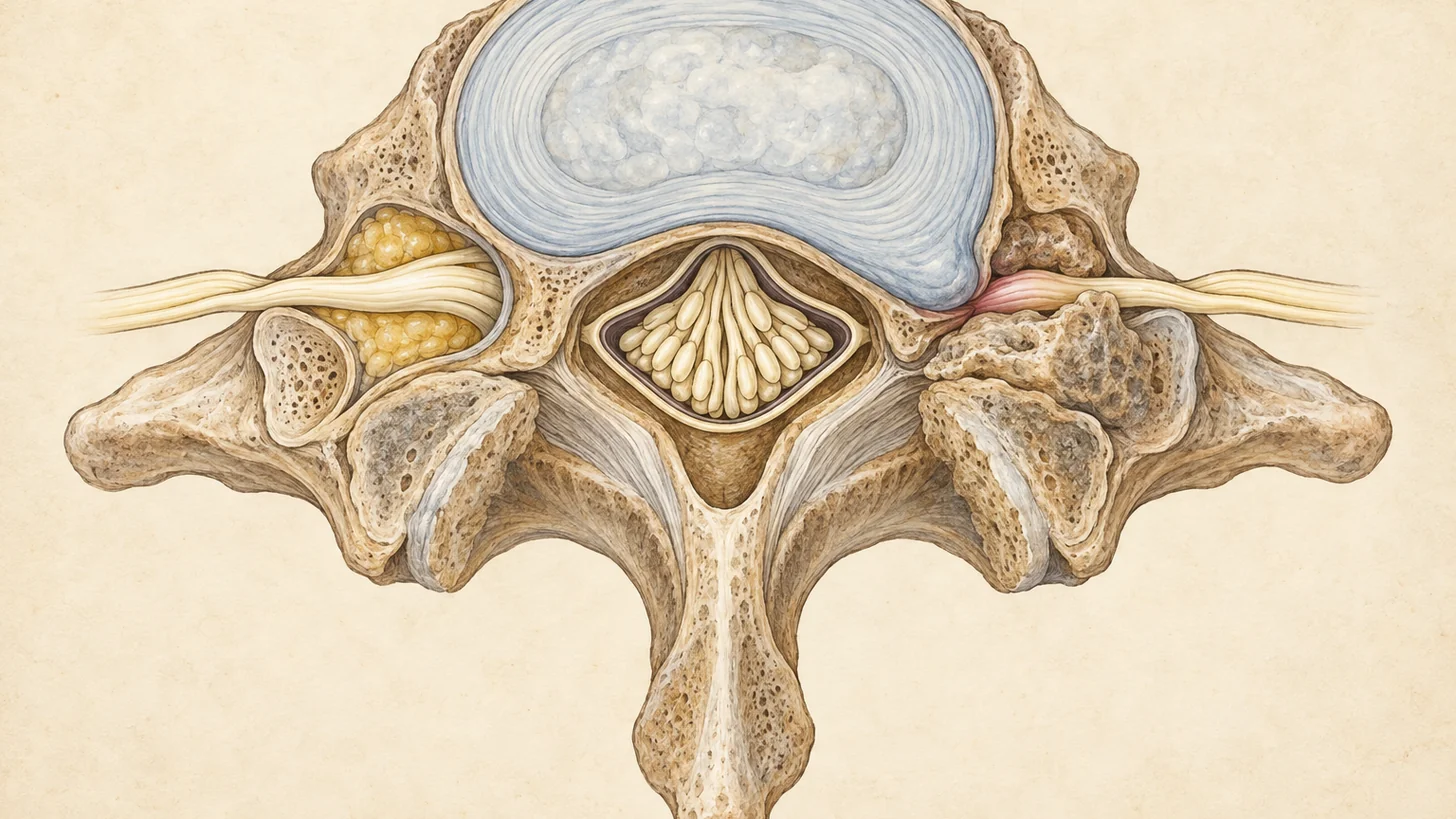
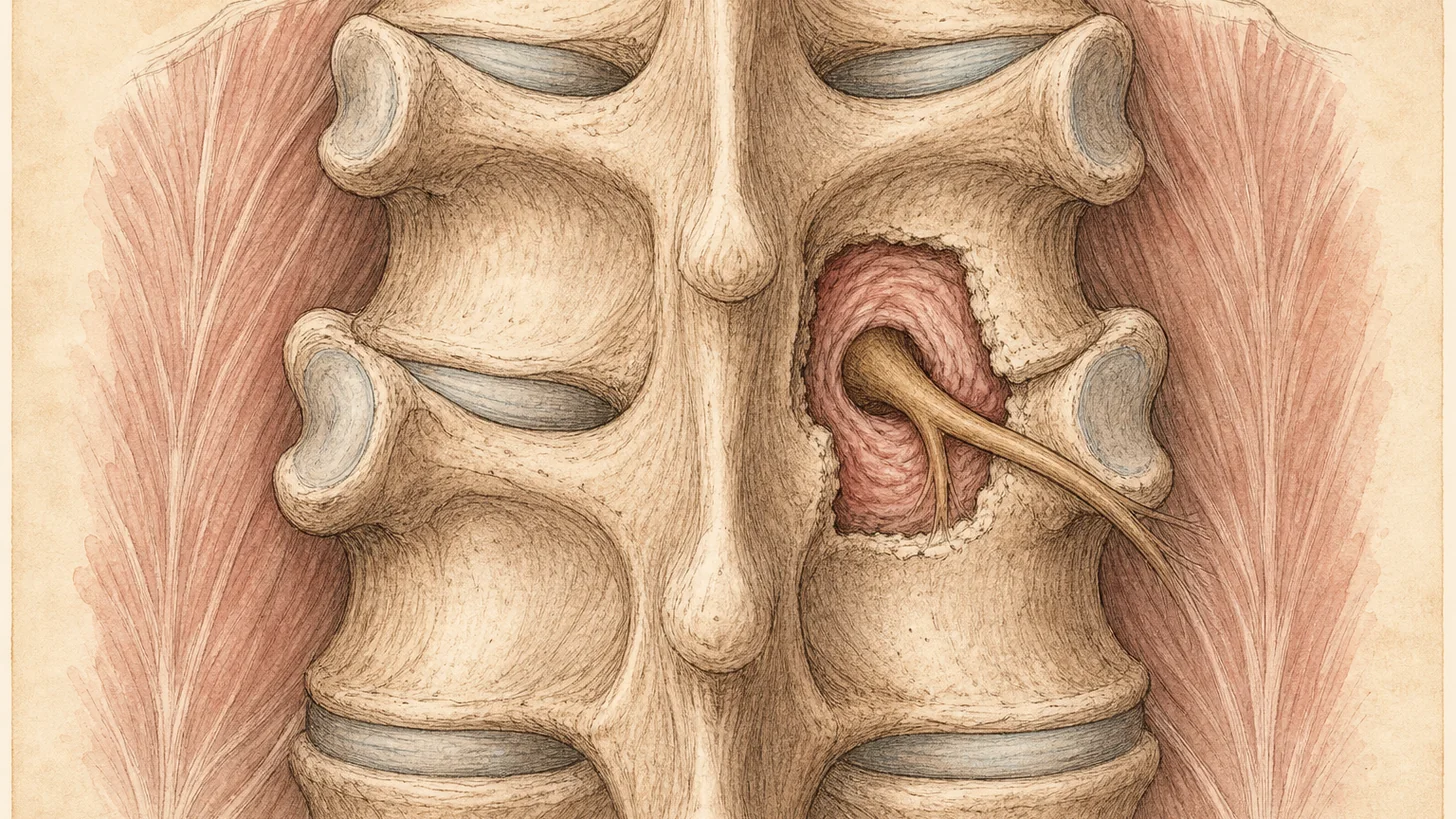
Spinal stenosis means a narrowing of the spaces inside the spine — the central canal that houses the spinal cord and cauda equina, or the lateral recesses and neural foramina where individual nerve roots exit. When these spaces narrow, the nerves within them are compressed, producing pain, numbness, tingling, and weakness in the territory those nerves supply. The condition is most often degenerative, developing gradually from the accumulated wear of decades. Less commonly it is congenital (a naturally narrow canal that becomes symptomatic with only mild degenerative change) or secondary to spondylolisthesis, trauma, or prior surgery. Authoritative overviews of the condition are published by the National Institute of Arthritis and Musculoskeletal and Skin Diseases (NIAMS) and the National Institute of Neurological Disorders and Stroke (NINDS).
Two points matter for treatment planning. First, the location of the narrowing determines the symptom pattern — lumbar stenosis affects the legs, cervical stenosis affects the arms and, in severe cases, the spinal cord itself. Second, the severity on imaging does not reliably predict the severity of symptoms. Some patients with marked narrowing on MRI have modest complaints, while others with moderate stenosis can barely walk a block. This is why treatment should target your functional limitation and pain, not the picture on the scan — a principle that guides every plan Dr. Movshis builds.
Lower back or neck: two conditions with one name
Lumbar stenosis narrows the canal below the end of the spinal cord, so it squeezes the bundle of nerve roots there (the cauda equina) and shows up in the legs. Its rare emergency is cauda equina syndrome. Cervical stenosis narrows the canal around the spinal cord itself. When it pinches a single root it causes arm pain, and when it affects the cord it causes clumsy hands and an unsteady walk (myelopathy), which needs a spine surgeon’s evaluation, not an injection. This page is about the lumbar spine. The neck is covered on the cervical stenosis page, including how the exam separates a pinched root from a compressed cord.
Lumbar stenosis is the more common of the two in people who come in for walking trouble. Patients describe a shrinking “walking budget”: they can walk to the corner, then must stop, and next month it is half a block. Many notice that pushing a shopping cart or leaning on a walker lets them go farther, because the flexed posture mechanically opens the canal.
Causes and Risk Factors
Degenerative lumbar spinal stenosis is rarely caused by a single structure. The canal narrows from several directions at once:
- Ligamentum flavum thickening. The ligament along the back wall of the canal hypertrophies and buckles inward with age — often the single largest contributor to central stenosis.
- Facet joint arthropathy. Arthritic enlargement of the facet joints narrows the lateral recess and foramen from behind. The same facet joints are frequently an independent pain generator, which is why stenosis and facet-mediated back pain so often coexist.
- Disc bulging. Degenerated discs lose height and bulge circumferentially, narrowing the canal from the front.
- Osteophytes (bone spurs). Arthritic bone overgrowth further encroaches on the canal and foramina.
- Degenerative spondylolisthesis. Slippage of one vertebra on another (most often L4 on L5) compounds the narrowing.
Risk factors include age over 50, a congenitally narrow canal, female sex (degenerative spondylolisthesis is more common in women), prior lumbar surgery, and conditions such as Paget’s disease or achondroplasia that alter bone architecture. A congenitally narrow canal explains why some patients develop stenosis symptoms in their 40s with only modest degenerative change — there was little room to spare to begin with.
Neurogenic claudication: the shrinking walking distance
The signature of lumbar spinal stenosis is neurogenic claudication — poorly localized pain, heaviness, cramping, numbness, or weakness in the buttocks, thighs, and legs that is provoked by standing upright and walking and relieved within minutes by sitting or bending forward. It differs from vascular claudication (poor circulation) in several clinically useful ways: vascular pain is typically a tight calf cramp at a reproducible walking distance that stops promptly with standing still, while neurogenic claudication often requires sitting or flexion for relief and may be accompanied by tingling or back pain. The Cleveland Clinic’s overview of spinal stenosis summarizes this symptom pattern well for patients.
Other common features include:
- Low back pain that coexists with the leg symptoms
- Symptoms that are positional — better flexed, worse extended (lying on your stomach or arching backward narrows the canal further)
- Numbness or “dead” feeling in the feet or legs after walking
- Subjective leg weakness or unsteadiness after prolonged standing
- Night symptoms or restless legs–type discomfort in some patients
In a city where the day runs on foot, the limit shows up as a route problem before it shows up as a pain problem: the walk from Grand Central to the office gets broken into stops, walking down the subway stairs gets harder than climbing them, and standing in a line hurts more than walking a block. Sitting down on a bench, or leaning on a railing, is often the giveaway, because both flex the lower spine.
When leg pain follows a single sharp nerve-root distribution — for example, from the buttock down the back of one leg to the foot — that pattern overlaps with sciatica, and a disc herniation or foraminal stenosis at one level may be the driver rather than multi-level central stenosis. Distinguishing these patterns on examination changes the injection strategy. If your report lists foraminal narrowing at several levels, our guide to foraminal stenosis on MRI explains what the grades mean and why most of them are silent.
Call 911 or go to the nearest emergency room if you develop new loss of bladder or bowel control, numbness in the groin or “saddle” area, or rapidly progressive weakness in both legs. These suggest cauda equina compression, which is a surgical emergency.
Diagnosis and Assessment
Diagnosis begins with the story. The most useful single findings are no pain when seated and relief bending forward (Suri 2010), and the examination then checks the nerves and rules out look-alikes. Dr. Movshis performs a focused neurological and musculoskeletal examination: gait observation (including whether symptoms can be provoked by treadmill-free walking in the office or by lumbar extension), strength, reflexes, and sensation testing in the legs, and provocative maneuvers to sort out how much of the pain is radicular (nerve-root) versus facet-mediated versus myofascial. A vascular screen — palpating foot pulses and reviewing risk factors — helps exclude peripheral arterial disease when the claudication story is ambiguous.
MRI of the lumbar spine without contrast is the standard confirmatory study: it shows the degree and levels of canal narrowing, ligamentum flavum thickness, disc contribution, and foraminal compromise. CT is reserved for patients who cannot have MRI or when fine bony detail matters, and flexion–extension X-rays assess stability when spondylolisthesis is suspected. Electrodiagnostic testing (EMG/nerve conduction studies) is occasionally useful to distinguish stenosis from peripheral neuropathy when the examination is equivocal. If you already have imaging, bring it — a prior MRI is frequently sufficient, and we review the actual images with you, not just the report.
One diagnostic tool specific to interventional practice: when it is unclear whether the leg symptoms or the back pain is the dominant problem, a diagnostic injection can help. A fluoroscopically guided medial branch block that temporarily relieves the back pain makes a facet contribution likely, and a selective nerve root block can point to which foramen is symptomatic. Neither proves the source, but both make the plan more specific than treating “stenosis” generically.
How lumbar stenosis is treated, step by step
There is no single “stenosis procedure” — there is a sequence, matched to your anatomy, your symptom pattern, and your response at each step. Our approach is layered, non-opioid, and evidence-based.
1. Conservative foundation
Structured physical therapy emphasizing flexion-biased exercises, hip and core strengthening, and activity modification is the evidence-supported starting point. NSAIDs can help symptomatically for appropriate patients. We are candid about the limits here: exercise cannot physically reopen a narrowed canal, and high-quality evidence for any non-surgical treatment of stenosis is lacking. A Cochrane review found low-quality evidence from a single trial that exercise gives short-term benefit for leg pain and function (Ammendolia 2013). In a randomized trial of surgical candidates, a physical therapy program improved physical function about as much as decompression at two years (Delitto 2015).
2. Epidural steroid injections (ESI)
Epidural steroid injections are the most commonly used interventional treatment for symptomatic lumbar stenosis, though not the best supported. Under fluoroscopic guidance, corticosteroid is delivered into the epidural space to reduce inflammation around the compressed nerve roots. For central stenosis with bilateral or multi-level symptoms, an interlaminar approach gives broad coverage. For single-level foraminal stenosis with one-sided radicular pain, a transforaminal approach targets the exiting root directly, and a caudal approach suits certain lower-canal patterns. We set expectations honestly. The LESS trial in the New England Journal of Medicine found that in 400 stenosis patients, adding steroid to epidural lidocaine made no significant difference in disability or leg pain at six weeks (Friedly 2014), and repeat injections added nothing long-term when the first six weeks had not helped (Friedly 2017). Pooled trials found no clear effect of epidural steroids in stenosis (Chou 2015). That is why we treat ESI as one tool in a plan, mainly for a flare or for one pinched root in a narrowed foramen, reassess response after each injection, and stop when it is not working.
3. Targeted nerve blocks
When a specific nerve root is the pain generator — common in foraminal stenosis — a selective nerve root block delivers anesthetic (with or without steroid) to that root, which can help identify it and can also treat it.
4. Treating the facet contribution
Because arthritic facet enlargement both narrows the canal and generates pain independently, many stenosis patients benefit from addressing the facets. A positive medial branch block (two blocks, each giving at least 80% relief) identifies candidates for radiofrequency ablation of the medial branch nerves, which can give months of back-pain relief in people who respond, though it does not change the canal diameter and does not work for everyone (Cohen 2020).
5. Referral-level options: MILD and surgery
Two options sit outside our office-based scope, and we discuss them candidly. The MILD procedure (percutaneous image-guided lumbar decompression) removes small portions of thickened ligamentum flavum through a port about the diameter of a pencil, under imaging guidance, without general anesthesia. It is designed specifically for ligamentum-flavum–dominant lumbar stenosis with neurogenic claudication. The main randomized trial compared it with epidural steroid injections rather than a sham, was unblinded and was sponsor-supported: at one year, 58% of the MILD group and 27% of the injection group met the disability-improvement target (Benyamin 2016). Modal Pain Management does not currently perform the MILD procedure in-house. When a patient’s anatomy and insurance profile make it a reasonable consideration, we refer to qualified interventional colleagues in Manhattan and coordinate care before and after. Similarly, when stenosis produces progressive neurological deficit, myelopathy, or refractory symptoms after a genuine trial of conservative and interventional care, decompressive surgery (laminectomy/laminotomy, with fusion in selected cases) is the appropriate path, and we make that referral without delay and without framing it as a failure.
Elective surgery for symptoms that are disabling but not progressing is a genuine choice, not an obligation. In the SPORT stenosis trial, decompression did better than non-surgical care on all primary outcomes in the as-treated analysis, while the intention-to-treat difference was limited to bodily pain (Weinstein 2008). In a randomized trial of 169 surgical candidates aged 50 or older, physical function improved by 22.4 points after decompression and 19.2 points after a physical therapy program at two years, a difference that was not significant, although 57% of the therapy group crossed over to surgery along the way (Delitto 2015). Some people want the operation and some never need it, and both can be the right decision.
What to Expect During Your Visit
Your first appointment is a consultation, not a procedure. Dr. Movshis reviews your history and prior imaging (bring a printed copy of your MRI report, or email your imaging to info@modalpain.com ahead of your visit), performs the examination described above, and explains — in plain language, with your images on screen — which structures are narrowing your canal and which are generating your pain. You will leave with a written plan: what we recommend first, why, what the alternatives are, and what response would trigger the next step.
If an epidural injection or nerve block is part of the plan, it is performed in our office procedure suite under fluoroscopic guidance. The skin is numbed with local anesthetic, and most patients feel pressure rather than pain. The injection itself takes roughly 15–20 minutes, followed by a short monitored recovery period. You should arrange a ride home. Most patients return to desk work the next day and avoid heavy lifting and strenuous exercise for 48 hours. We typically schedule a follow-up at two to four weeks to grade the response and decide whether a second injection, a different target, or a different strategy is indicated.
Cost and Insurance for Spinal Stenosis Treatment
Modal Pain Management accepts most commercial PPO plans, including Aetna, Anthem Blue Cross Blue Shield, Cigna, Empire Blue Cross Blue Shield, the Empire Plan, Oscar, Oxford, UMR and UnitedHealthcare. We check your benefits and go over what your plan covers and any expected cost (deductible, coinsurance, and copay) by phone before your visit, at no cost or obligation, so there are no surprise bills. With commercial PPO coverage, interventional spine procedures such as epidural steroid injections are typically covered benefits when medically indicated, and what you owe depends on your plan’s deductible and coinsurance. When your plan requires prior authorization, our office submits it after the consultation.
We do not accept Medicare, Medicaid, HMO plans, or workers’ compensation. If you carry one of these, we will tell you directly rather than book you into a visit you cannot use. Transparent self-pay pricing is available for patients without usable coverage, and insurance verification takes one business day in most cases. We do not bill surprise facility fees, because procedures are performed in our office suite, not a hospital outpatient department.
Self-pay prices are flat and cover the physician fee, the fluoroscopic guidance, and the medication:
- New-patient consultation: $250
- Lower-back epidural steroid injection, interlaminar or caudal: $1,000
- Lumbar transforaminal epidural: $1,000 for the first level, plus $500 for each additional level in the same session
- Lower-back medial branch block: $1,000
- Lower-back radiofrequency ablation, up to three levels: $1,500
HSA and FSA cards are accepted, and payment plans are available through Cherry.
Recovery and Long-Term Management
Lumbar spinal stenosis is a chronic structural condition, and honest long-term management means planning for that. After an injection, relief from the anesthetic is immediate but temporary, while the steroid works over days, and the result is judged two to four weeks later. In stenosis the benefit is often small or short, because the mechanical narrowing remains (Friedly 2014). That is the rationale for pairing any procedure with sustained exercise and activity strategies rather than relying on injections.
Many patients do well on a maintenance rhythm — periodic reassessment, a repeat epidural injection when symptoms rebuild (within safe annual limits), and continuous home exercise. Symptoms often wax and wane, and some patients stay stable for long periods (Lurie 2016). A smaller group progresses despite appropriate care, and for them we facilitate surgical consultation rather than escalating injections indefinitely. Our commitment is to the trajectory of your function — how far you can walk, how well you sleep, whether you can travel and work — not to any single procedure.
Why Choose Modal Pain Management for Spinal Stenosis?
- Specialist evaluation, not protocol care. Your evaluation and every procedure are performed by Dr. Movshis, a Mount Sinai fellowship-trained, dual board-certified interventional pain physician — not delegated to extenders.
- Image-guided, office-based care. All injections are performed under fluoroscopy in our suite at 369 Lexington Ave, Floor 25 — no hospital referral, no facility fees, same-week scheduling.
- Non-opioid by design. Treatment plans are built around interventional and rehabilitative care. Modal Pain does not prescribe opioids.
- Honest triage. When your imaging and examination point past injections — to MILD or to surgery — we say so and coordinate the referral.
- Midtown convenience. Minutes from Grand Central, serving Midtown East, Murray Hill, Turtle Bay, Kips Bay, and Gramercy. See our Midtown service area page for directions.
References
- Lurie J, Tomkins-Lane C. Management of lumbar spinal stenosis. BMJ. 2016;352:h6234. doi:10.1136/bmj.h6234 · PubMed
- Ammendolia C, Stuber KJ, Rok E, et al. Nonoperative treatment for lumbar spinal stenosis with neurogenic claudication. Cochrane Database Syst Rev. 2013;2013(8):CD010712. doi:10.1002/14651858.CD010712 · PubMed
- Delitto A, Piva SR, Moore CG, et al. Surgery versus nonsurgical treatment of lumbar spinal stenosis: a randomized trial. Ann Intern Med. 2015;162(7):465–73. doi:10.7326/M14-1420 · PubMed
- Friedly JL, Comstock BA, Turner JA, et al. A randomized trial of epidural glucocorticoid injections for spinal stenosis. N Engl J Med. 2014;371(1):11–21. doi:10.1056/NEJMoa1313265 · PubMed
- Chou R, Hashimoto R, Friedly J, et al. Epidural Corticosteroid Injections for Radiculopathy and Spinal Stenosis: A Systematic Review and Meta-analysis. Ann Intern Med. 2015;163(5):373–81. doi:10.7326/M15-0934 · PubMed
- Benyamin RM, Staats PS, MiDAS Encore I. MILD® Is an Effective Treatment for Lumbar Spinal Stenosis with Neurogenic Claudication: MiDAS ENCORE Randomized Controlled Trial. Pain Physician. 2016;19(4):229–42. PubMed
- Cohen SP, Bhaskar A, Bhatia A, et al. Consensus practice guidelines on interventions for lumbar facet joint pain from a multispecialty, international working group. Reg Anesth Pain Med. 2020;45(6):424–467. doi:10.1136/rapm-2019-101243 · PubMed
- Centers for Medicare & Medicaid Services. Local Coverage Determination L39036: Epidural Steroid Injections for Pain Management. Effective May 7, 2026. cms.gov
- Friedly JL, Comstock BA, Turner JA, et al. Long-term effects of repeated injections of local anesthetic with or without corticosteroid for lumbar spinal stenosis: a randomized trial. Arch Phys Med Rehabil. 2017. doi:10.1016/j.apmr.2017.02.029 · PubMed
- Suri P, Rainville J, Kalichman L, Katz JN. Does this older adult with lower extremity pain have the clinical syndrome of lumbar spinal stenosis? JAMA. 2010;304(23):2628–36. doi:10.1001/jama.2010.1833 · PubMed
- Weinstein JN, Tosteson TD, Lurie JD, et al. Surgical versus nonsurgical therapy for lumbar spinal stenosis. N Engl J Med. 2008;358(8):794–810. doi:10.1056/NEJMoa0707136 · PubMed