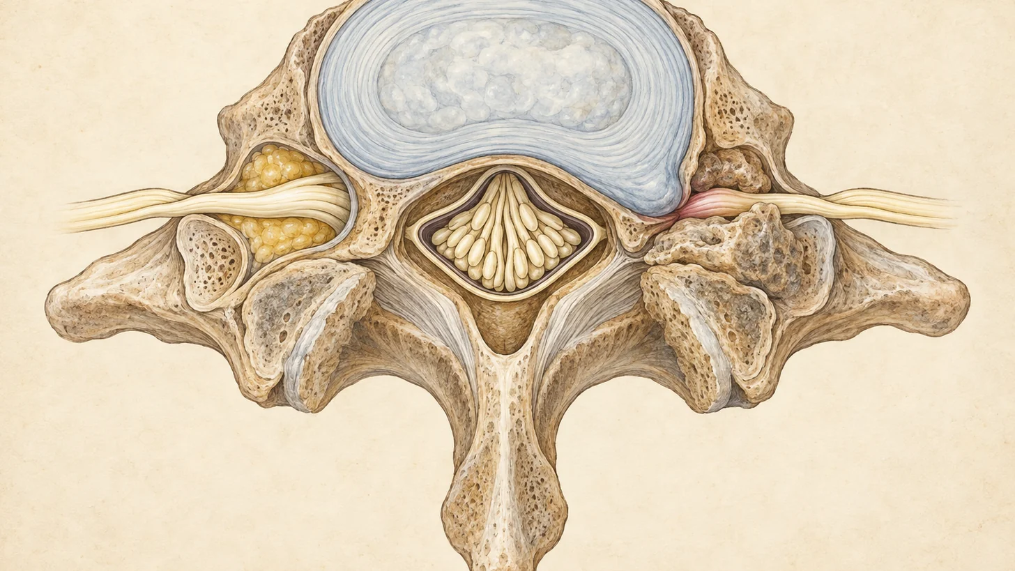
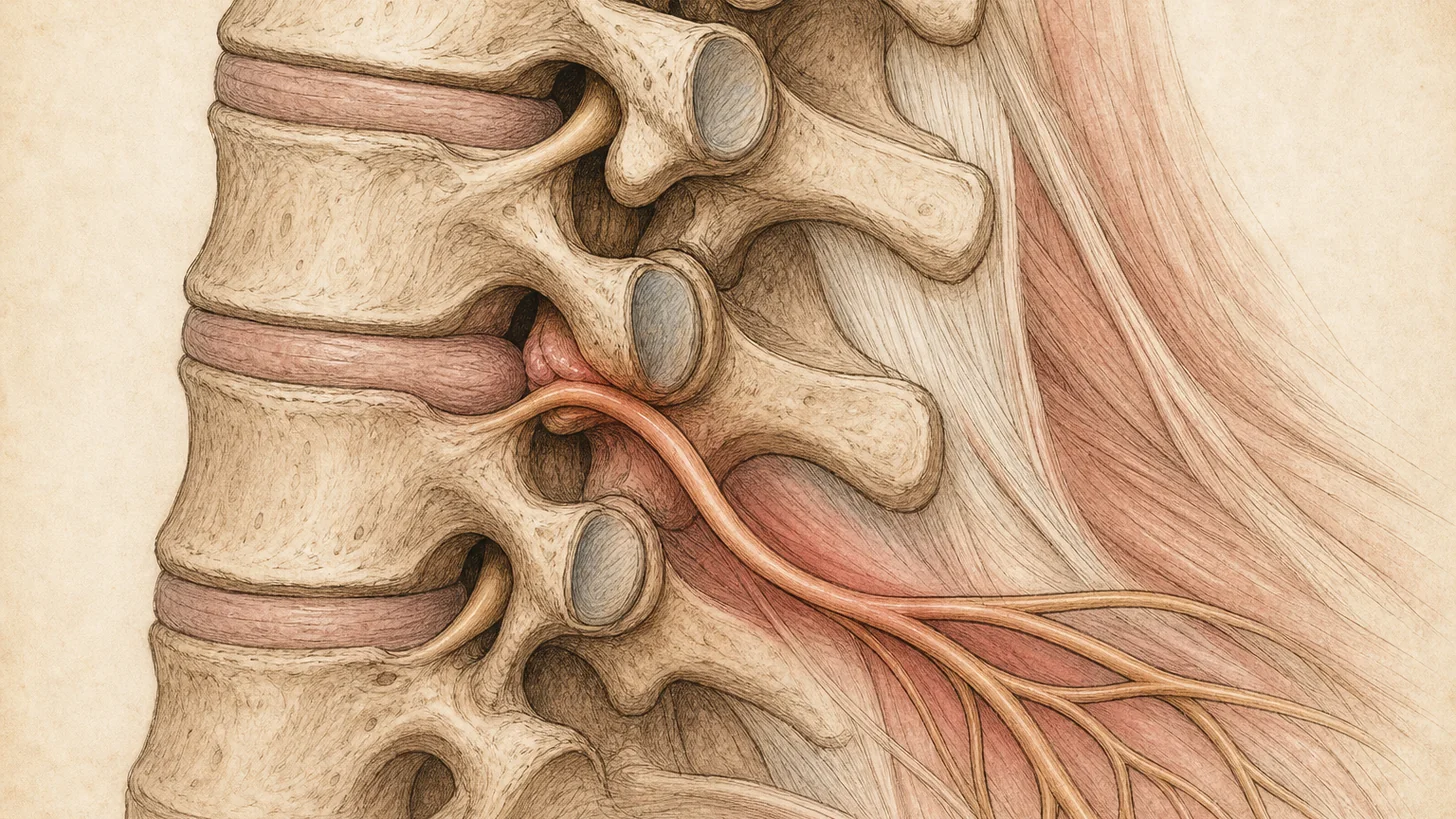
A 54-year-old litigator brings in an MRI report that reads: multilevel spondylosis, moderate central canal stenosis at C5–6 with mild flattening of the cord, severe left neural foraminal narrowing at C6–7. What brought her in is an ache running down the back of her left arm into her middle finger, worse when she looks up at a high monitor, better when she rests her hand on top of her head. She buttons her blouse without trouble, her handwriting has not changed, and she walks a straight line. That report describes two different problems. Only one of them is causing her pain, and only one of them would ever need a surgeon.
Cervical stenosis means narrowing inside the neck part of the spine. The narrowing comes from the same slow changes that gray the hair: discs lose height and bulge, the edges of the vertebrae grow bone spurs, the ligament along the back of the canal thickens and buckles, and the small joints enlarge. Where the narrowing sits decides what it does. At Modal Pain Management, at 369 Lexington Avenue, Floor 25, a short walk from Grand Central, Dr. Alex Movshis, MD, dual board-certified in anesthesiology and pain medicine, sorts that out on the first visit with an exam read against your imaging.
Three findings share one name on the report
Central canal stenosis. The tube that holds the spinal cord is narrowed, usually from the front by disc and bone spur and from behind by thickened ligament. A widely used MRI scale grades it from 0 to 3. Grade 1 means more than half of the fluid cushion around the cord is gone. Grade 2 means the cord itself is deformed, which is what radiologists mean by “flattening” or “effacement.” Grade 3 means a bright signal change inside the cord (Kang 2011). A narrow canal can be present for years without symptoms, because the cord tolerates a surprising amount of crowding until it does not.
Foraminal stenosis. Each nerve root leaves the spine through a side opening called the foramen, and arthritic joints and spurs at its edges close it down. The root that is squeezed belongs to the level below the disc number: C5–6 narrows the C6 root, C6–7 the C7 root. This is the stenosis that causes arm pain. Most reports grade it separately on each side, and a severe grade on the side and level that match your symptoms is meaningful. A severe grade on the other side, with no symptoms there, usually is not. Our guide to foraminal stenosis on MRI explains the grades and the left, right, and bilateral wording.
Cord compression or cord signal change. These phrases describe the cord being touched or altered on the scan. They raise the question of myelopathy, and they do not answer it.
The MRI finds more stenosis than there are patients
In 1,211 volunteers aged 20 to 70 with no neck or arm symptoms, 87.6% had disc bulging on cervical MRI and 5.3% had spinal cord compression they knew nothing about. Cord compression became more common after age 50 and sat mostly at C5–6 and C6–7 (Nakashima 2015), the same levels that show up on symptomatic patients’ reports. The scan cannot tell you which of those findings is yours.
The relationship runs both ways for the foramen. In a study that graded cervical foraminal narrowing on oblique MRI and compared it with a neurosurgeon’s exam, the two highest grades almost always came with matching nerve findings, yet 17% to 18% of patients whose foramina were graded normal still had nerve findings on exam (Park 2014). On the foramen, a normal-looking scan can miss a root that the exam finds.
Position matters too. Standard MRI is taken with the head neutral. When patients with cervical spondylosis were scanned in flexion and extension, the canal narrowed further in extension in 48% of them, and cord compression was seen in extension in 20%, against 11% in flexion (Muhle 1998). That is why looking up at a high monitor, painting a ceiling, or reaching overhead often brings on the arm symptoms, and why tipping the head back during the exam can reproduce them.
Root or cord: the exam decides
A pinched root produces pain that travels. It runs down one arm in a strip, toward the thumb and index finger for C6, the middle finger for C7, the ring and little fingers for C8, with tingling in the same strip and sometimes weakness in the muscles that root supplies. The reflex at that level may be reduced. Tipping the head back and toward the painful side reproduces it (the Spurling maneuver), and resting the hand on top of the head often relieves it. The level-by-level pattern, and how most of these settle, is laid out in our guide to a pinched nerve in the neck, and the full workup is on the radiculopathy page.
A compressed cord behaves differently, and it can be painless. The hands become clumsy: buttons, keys, typing on a phone, handwriting that gets worse. Both hands may feel numb in a way that does not follow one strip. Walking becomes unsteady, especially in the dark or on stairs. Some patients feel an electric jolt down the spine when they bend the neck forward. On exam, the reflexes are brisk rather than reduced. A finger flick that makes the thumb twitch (Hoffmann sign), a toe that goes up instead of down (Babinski sign), and a wide or wobbly gait are the findings. In 249 patients with cervical spine problems, a cluster of five findings, gait deviation, a positive Hoffmann sign, an inverted supinator sign, a positive Babinski sign, and age over 45, performed well as a set: three or more of the five raised the odds of myelopathy about 31-fold, and none of the five cut them to less than a fifth (Cook 2010). That cluster is part of the first-visit exam for anyone whose report mentions the cord.
Many patients have both. A root pinched at C6–7 and a canal narrowed at C5–6 can sit one level apart, as in the litigator above. Her arm pain came from the C7 root. Her normal hands, normal gait, and normal reflexes meant the cord, though crowded on the scan, was not yet affected.
Go to the closest emergency room or call 911 for weakness in the arms or legs that is getting worse by the day, new trouble controlling your bladder or bowel, or new numbness or weakness after a fall or a blow to the head or neck. A canal that is already narrow leaves the cord less room when the neck is forced backward.
Cord compression with a normal exam
This is the finding that frightens people most, and it is the one where the evidence is clearest about restraint. In a systematic review of patients with cervical canal stenosis and cord compression from spondylosis but no myelopathy, about 8% developed myelopathy at one year and 23% over a median of 44 months (Wilson 2013). Most did not. The 2017 AOSpine guideline suggests not offering preventive surgery to these patients when they have no root symptoms either. It suggests that they be told the risk, taught the warning signs, and followed clinically. Patients whose compression comes with radiculopathy are at higher risk of progressing, and the guideline suggests offering them either surgery or nonsurgical care with close follow-up (Fehlings 2017).
In practice that means a documented baseline exam, so a later exam has something to be compared against, a clear list of the signs that should bring you back sooner, and regular re-examination. When arm symptoms from a pinched root sit on top of cord compression, a surgical opinion is offered alongside the nonsurgical plan, so the decision is made with both options on the table.
When the narrowing pinches a nerve root
Cervical radiculopathy is a disease of middle age. In a population study from Rochester, Minnesota, the annual incidence peaked at ages 50 to 54, and 68.4% of cases were related to spondylosis, disc, or both. Over a median follow-up of almost five years, 31.7% had a recurrence and 26% had surgery, and at last follow-up 90% were symptom-free or only mildly limited (Radhakrishnan 1994).
For spondylotic root compression, surgery mostly buys speed. In a randomized trial of 81 patients whose root was clearly compressed on MRI or CT myelography, the surgery group had less pain, better strength, and less numbness at four months. A year after that, pain, numbness, and tingling were the same whether patients had surgery, physical therapy, or a collar, although the surgery group kept an edge in some strength measures over physical therapy (Persson 1997). That is the case for a structured nonsurgical plan first, with surgery kept for weakness that progresses or pain that does not respond.
Treatment follows the root-or-cord answer
Arm pain from a narrowed foramen
Physical therapy is the base, in our own office at 369 Lexington: posture and loading changes, deep neck flexor and scapular strengthening, and nerve mobility work, with the head-back positions that narrow the foramen trimmed out of the day. Most pinched roots settle over weeks to months.
When the arm pain is keeping you from sleeping or working, or has not eased after several weeks, the next step is a fluoroscopically guided cervical epidural steroid injection. It calms the inflammation around the pinched root. It does not widen the foramen, and it is paired with therapy rather than replacing it. In a randomized trial of 169 patients with cervical radicular pain, arm pain at one month did not differ among the injection, medication with physical therapy, and the combination, but at three months 56.9% of the combination group had a positive outcome, against 36.7% with the injection alone and 26.8% with medication and therapy alone (Cohen 2014).
Safety in a narrow canal is about where the needle goes and what goes through it. National safety recommendations place a cervical interlaminar injection at C7–T1 ideally and no higher than C6–7, where the space behind the dura is widest (Rathmell 2015). Stenosis is exactly what thins that space, so the MRI is reviewed for the size of the epidural space at the entry level before every cervical injection, as the same recommendations advise (Rathmell 2015). Dr. Movshis uses the interlaminar route in the neck, under live fluoroscopy with contrast, and dexamethasone, a non-particulate steroid. In a systematic review, non-particulate steroid relieved cervical pain about as well as particulate steroid (Mehta 2017), and particulate steroid entering an artery is the mechanism blamed for the rare strokes and spinal cord injuries reported after cervical transforaminal injections (Rathmell 2015). If a well-placed injection does nothing, it is not repeated at the same level in hope.
Neck pain from arthritic facet joints
The same arthritis that narrows the foramen enlarges the facet joints behind it, and those joints hurt on their own: deep, one-sided neck pain, worse tipping the head back or turning, with no arm symptoms. The MRI cannot say whether a facet joint is the source, because facet arthritis is common on the MRIs of people with no neck pain at all. A cervical medial branch block tests it, and radiofrequency ablation of the same small nerves treats it when the block confirms. The neck pain page covers how the four common neck pain sources are told apart.
Cervical myelopathy
Injections do not decompress a spinal cord, and they are not used to treat myelopathy. The guideline group grades severity with the modified Japanese Orthopaedic Association score: 15 to 17 is mild, 12 to 14 moderate, and 11 or less severe (Fehlings 2017). For moderate and severe myelopathy it recommends surgery. For mild myelopathy it suggests either surgery or a supervised trial of structured rehabilitation, with surgery if the patient worsens or fails to improve (Fehlings 2017).
Dr. Movshis does not perform surgery. When the exam shows myelopathy, the referral to a spine surgeon happens that week, and the letter carries what the surgeon needs to act on it: the hand, gait, and reflex findings, the score, the MRI levels, and anything that has been tried. Patients with mild myelopathy who choose a monitored nonsurgical course are re-examined against that baseline, so a decline is caught by measurement rather than by the patient noticing it late.
What cervical stenosis care costs
Most commercial PPO plans cover the consultation and the procedures on this page, including Aetna, Anthem Blue Cross Blue Shield, Cigna, Empire Blue Cross Blue Shield, the Empire Plan, Oscar, Oxford, UMR, and UnitedHealthcare. When a plan requires prior authorization for an epidural injection or a medial branch block, our office submits it after the consultation. Modal Pain Management does not accept Medicare, Medicaid, HMO plans, or workers’ compensation. You can verify your insurance before the visit.
Self-pay prices are flat and cover the physician fee, the fluoroscopic guidance, and the medication, with no separate facility fee:
- New-patient consultation: $250
- Cervical epidural steroid injection: $1,500
- Cervical medial branch block: $1,500
- Cervical radiofrequency ablation, up to three levels: $2,000
HSA and FSA cards are accepted, and payment plans are available through Cherry. Every price is on the self-pay pricing page.
The first visit
The consultation is 45 minutes. Bring the MRI images, not only the report, and a list of what has been tried. Dr. Movshis takes the history, examines the neck, both arms, the hands, the reflexes, and your walking, and then reads the imaging against what the exam found, with the images on screen. You leave knowing whether the narrowing is pinching a root, affecting the cord, both, or neither, and what the next step is. Procedures are typically scheduled within one to two weeks in the same office, take 15 to 30 minutes, and you go home the same day with a driver. If you are not sure the problem is in your neck at all, the Pain Self-Assessment takes the history a pain physician takes and gives you a printable summary for any doctor you see.
When the narrowing is in your lower back and your legs give out on long walks instead, that is lumbar stenosis, a different condition with a different plan, covered on our spinal stenosis page.
References
- Fehlings MG, Tetreault LA, Riew KD, et al. A Clinical Practice Guideline for the Management of Patients With Degenerative Cervical Myelopathy: Recommendations for Patients With Mild, Moderate, and Severe Disease and Nonmyelopathic Patients With Evidence of Cord Compression. Global Spine J. 2017;7(3 Suppl):70S–83S. doi:10.1177/2192568217701914 · PubMed
- Fehlings MG, Tetreault LA, Riew KD, Middleton JW, Wang JC. A Clinical Practice Guideline for the Management of Degenerative Cervical Myelopathy: Introduction, Rationale, and Scope. Global Spine J. 2017;7(3 Suppl):21S–27S. doi:10.1177/2192568217703088 · PubMed
- Wilson JR, Barry S, Fischer DJ, et al. Frequency, timing, and predictors of neurological dysfunction in the nonmyelopathic patient with cervical spinal cord compression, canal stenosis, and/or ossification of the posterior longitudinal ligament. Spine (Phila Pa 1976). 2013;38(22 Suppl 1):S37–54. doi:10.1097/BRS.0b013e3182a7f2e7 · PubMed
- Nakashima H, Yukawa Y, Suda K, et al. Abnormal findings on magnetic resonance images of the cervical spines in 1211 asymptomatic subjects. Spine (Phila Pa 1976). 2015;40(6):392–8. doi:10.1097/BRS.0000000000000775 · PubMed
- Kang Y, Lee JW, Koh YH, et al. New MRI grading system for the cervical canal stenosis. AJR Am J Roentgenol. 2011;197(1):W134–40. doi:10.2214/AJR.10.5560 · PubMed
- Park HJ, Kim SS, Han CH, et al. The clinical correlation of a new practical MRI method for grading cervical neural foraminal stenosis based on oblique sagittal images. AJR Am J Roentgenol. 2014;203(2):412–7. doi:10.2214/AJR.13.11647 · PubMed
- Muhle C, Weinert D, Falliner A, et al. Dynamic changes of the spinal canal in patients with cervical spondylosis at flexion and extension using magnetic resonance imaging. Invest Radiol. 1998;33(8):444–9. doi:10.1097/00004424-199808000-00004 · PubMed
- Cook C, Brown C, Isaacs R, et al. Clustered clinical findings for diagnosis of cervical spine myelopathy. J Man Manip Ther. 2010;18(4):175–80. doi:10.1179/106698110X12804993427045 · PubMed
- Radhakrishnan K, Litchy WJ, O’Fallon WM, Kurland LT. Epidemiology of cervical radiculopathy. A population-based study from Rochester, Minnesota, 1976 through 1990. Brain. 1994;117(Pt 2):325–35. doi:10.1093/brain/117.2.325 · PubMed
- Persson LC, Moritz U, Brandt L, Carlsson CA. Cervical radiculopathy: pain, muscle weakness and sensory loss in patients with cervical radiculopathy treated with surgery, physiotherapy or cervical collar. A prospective, controlled study. Eur Spine J. 1997;6(4):256–66. doi:10.1007/BF01322448 · PubMed
- Cohen SP, Hayek S, Semenov Y, et al. Epidural steroid injections, conservative treatment, or combination treatment for cervical radicular pain: a multicenter, randomized, comparative-effectiveness study. Anesthesiology. 2014;121(5):1045–55. doi:10.1097/ALN.0000000000000409 · PubMed
- Rathmell JP, Benzon HT, Dreyfuss P, et al. Safeguards to prevent neurologic complications after epidural steroid injections: consensus opinions from a multidisciplinary working group and national organizations. Anesthesiology. 2015;122(5):974–84. doi:10.1097/ALN.0000000000000614 · PubMed
- Mehta P, Syrop I, Singh JR, Kirschner J. Systematic Review of the Efficacy of Particulate Versus Nonparticulate Corticosteroids in Epidural Injections. PM R. 2017;9(5):502–512. doi:10.1016/j.pmrj.2016.11.008 · PubMed