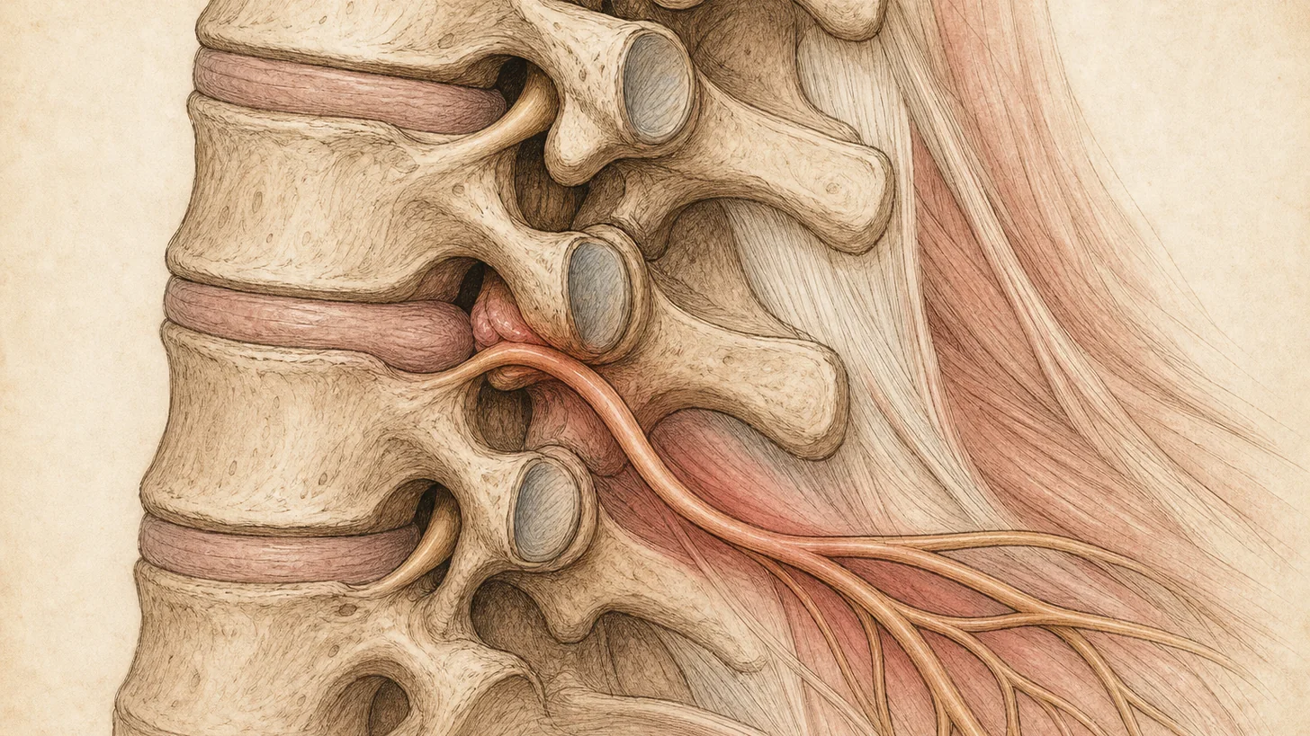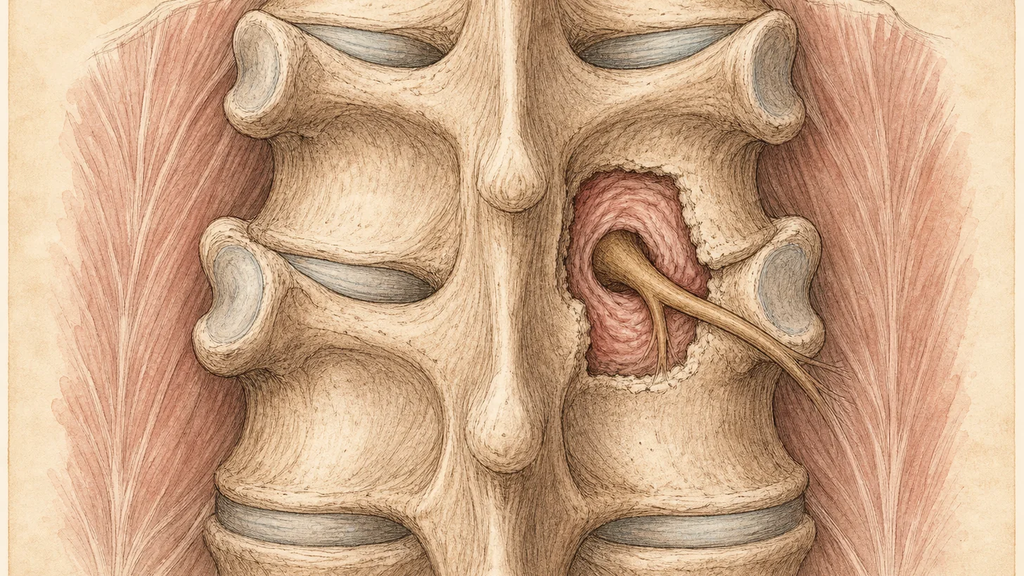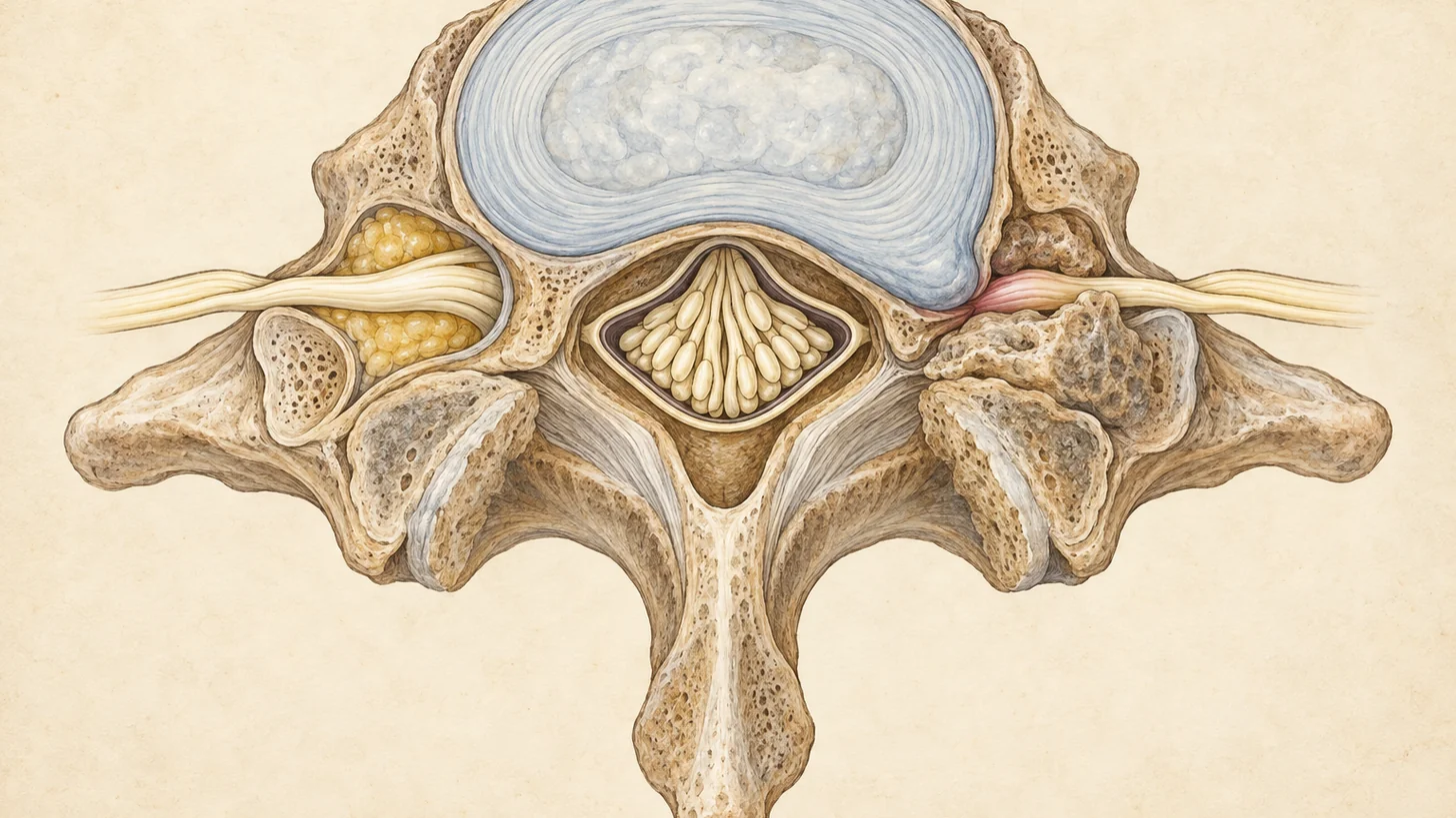
A 61-year-old brings in a lumbar MRI report: moderate bilateral neural foraminal narrowing at L4–5, severe left neural foraminal stenosis at L5–S1. He has read it a dozen times. His pain runs down the back of his right calf to the outside of his right foot. The word “severe” is on the left. That mismatch is the most useful thing on the page, and it is the kind of detail that gets lost when a report is read without an exam.
What the foramen is, and what closes it
Every level of the spine has two foramina, one on each side. Each is a short bony tunnel bounded by the vertebra above and below, the disc in front, and the small facet joint behind. A single nerve root runs through it, cushioned by fat and accompanied by small blood vessels.
The tunnel narrows from several directions at once, which is why the finding is so common with age. The disc loses height, so the roof drops. The disc bulges backward into the front of the opening. Bone spurs grow from the vertebral edges, and in the neck from the small uncovertebral joints. The facet joint enlarges with arthritis and closes the opening from behind. A slipped vertebra (spondylolisthesis) can pinch it further. None of this happens quickly, and most of it causes no symptoms.
Which nerve each level squeezes
The root inside a foramen is the one that exits there, and this is where reports confuse people.
In the neck, the root is numbered for the vertebra below the disc. The C5–6 foramen holds the C6 root (pain toward the thumb and index finger), C6–7 holds C7 (the middle finger), and C7–T1 holds C8 (the ring and little fingers). The level-by-level detail, with the muscles and reflexes each root controls, is in our guide to a pinched nerve in the neck.
In the lower back, the root is numbered for the vertebra above. The L4–5 foramen holds the L4 root, which runs to the front of the thigh, the knee, and the inner shin. The L5–S1 foramen holds the L5 root, which runs down the outer shin to the top of the foot and the big toe. There is a twist that matters for reading a report: a disc bulge at L4–5 that sits toward the middle of the canal presses on the L5 root on its way down, not the L4 root that exits beside it. “L4–5” on a report can therefore mean either root, depending on where the narrowing sits.
Back to the 61-year-old. Pain in the back of the calf and the outside of the foot follows S1, not L5, and it is on the right, not the left. The severe left L5–S1 foraminal stenosis does not explain it. Something else does, and the exam is how you find it.
What mild, moderate, and severe mean
Many radiologists grade by eye, but a widely used lumbar scale [1] defines the words in a way that is worth knowing:
- Mild: the fat around the nerve root is obliterated in two opposite directions, top and bottom or front and back. The root has less room but keeps its normal shape.
- Moderate: the fat is obliterated all the way around, in all four directions, but the root still has its normal shape.
- Severe: the root itself is collapsed or deformed.
Cervical foramina are graded on a similar scale. When graded cervical foramina were compared with a neurosurgeon’s exam, the two highest grades almost always came with matching nerve findings. Yet 17% to 18% of patients whose foramina were graded normal still had nerve findings on exam [2]. Severe narrowing on the symptomatic side is meaningful. A normal-looking foramen does not fully clear a root that the exam implicates.
Why the MRI calls more stenosis than there is
In a study of 90 surgical patients whose symptomatic level was confirmed by targeted decompression surgery, MRI was very good at ruling foramina out and poor at ruling them in. Across 936 foramina, MRI picked up 96% of the truly symptomatic ones, but only about 4% of the foramina that MRI called narrowed turned out to be the culprit [3]. A normal-looking opening on MRI is reassuring. A narrowed one is a candidate, not a diagnosis.
The wider picture is the same. Disc bulges, one of the main causes of foraminal narrowing, were found in 30% of pain-free 20-year-olds and 84% of pain-free 80-year-olds in a systematic review of spine imaging [4]. Age-related narrowing on a report is common and often silent.
“Bilateral” belongs in the same category. Both openings at a level share the same disc and the same pair of joints, so they tend to narrow together. Pain on one side is explained, if at all, by the opening on that side.
How a symptomatic foramen behaves
The pattern is one-sided and follows one nerve. In the lower back that means pain, burning, or numbness down one leg along a single root’s path, often worse standing, walking, or leaning backward, because extension narrows the foramen. In the neck it means pain down one arm toward particular fingers, often worse looking up, and better with the hand resting on top of the head.
Lumbar foraminal stenosis can also hurt at rest, which separates it from central canal stenosis, where the legs usually settle within minutes of sitting. In a prospective series of 30 patients with confirmed symptomatic foraminal stenosis, 20 had a swollen spinal nerve on MR myelography, and 95% of those 20 had leg pain at rest [5].
Central canal stenosis looks different: both legs grow heavy or numb after a few blocks, and sitting or leaning on a shopping cart brings relief within minutes. That pattern is covered on our spinal stenosis page. In the neck, narrowing that presses on the spinal cord rather than one root causes clumsy hands and an unsteady walk, and that needs a different conversation, covered on the cervical stenosis page.
Go to the closest emergency room or call 911 for leg or arm weakness that is getting worse by the day, new numbness in the groin or inner thighs, or new trouble controlling your bladder or bowel.
What happens next: match first, then treat
The first visit is the matching step. Dr. Movshis examines strength, reflexes, sensation, and the provocation tests for each root, then reads the MRI against what the exam found. In the lower back, when two levels are both plausible, a diagnostic selective nerve root block at one of them can settle which is responsible: if numbing that one root takes away the familiar leg pain, it is the right one. If nothing on the scan fits, the search moves to the hip, the sacroiliac joint, or a nerve trapped farther out in the limb.
When a foramen is the source, treatment follows a sequence:
- Physical therapy first, in our own office, built around keeping the spine out of the positions that narrow the opening and restoring the strength the pain took away.
- A transforaminal epidural steroid injection in the lower back, placed under fluoroscopy right next to the pinched root at that level. In a blinded trial in lumbar radicular pain, 54% of patients given steroid had at least half their pain gone at one month, against 7% to 21% with control injections [7]. In foraminal stenosis specifically, a retrospective study of 128 patients found pain and walking distance improved four weeks after the injection, with no control group, with better pain relief in moderate than in severe narrowing [6]. In the neck, Dr. Movshis uses the interlaminar route at a lower cervical level instead, for safety reasons explained on the epidural steroid injection page.
- A surgical opinion when the nerve is losing strength, or when the pain persists despite therapy and well-placed injections. Surgery that opens the foramen (foraminotomy) is a spine surgeon’s procedure, and Dr. Movshis refers with the exam and injection results attached.
Expect honesty about the limits. The injection calms inflammation around the root. It does not widen the opening. And the evidence is weaker when the problem is central canal stenosis rather than one pinched root: in a 400-patient double-blind trial in central stenosis, adding steroid to lidocaine made no significant difference at six weeks [8].
Self-pay prices are flat: $250 for the consultation, $1,000 for a lumbar transforaminal epidural at one level plus $500 for each additional level, and $1,500 for a cervical epidural. Every price is on the self-pay pricing page, and you can verify your insurance before the visit.
To book, call (646) 290-6660 or book online, and email the MRI images and report to info@modalpain.com before the visit.
References
This article is reviewed against the peer-reviewed literature. Citations retrieved from PubMed.
- Lee S, Lee JW, Yeom JS, et al.. A practical MRI grading system for lumbar foraminal stenosis. AJR. American Journal of Roentgenology. 2010. doi:10.2214/AJR.09.2772 · PubMed
- Park HJ, Kim SS, Han CH, et al.. The clinical correlation of a new practical MRI method for grading cervical neural foraminal stenosis based on oblique sagittal images. AJR. American Journal of Roentgenology. 2014. doi:10.2214/AJR.13.11647 · PubMed
- Aota Y, Niwa T, Yoshikawa K, Fujiwara A, Asada T, Saito T. Magnetic resonance imaging and magnetic resonance myelography in the presurgical diagnosis of lumbar foraminal stenosis. Spine. 2007. doi:10.1097/01.brs.0000259809.75760.d5 · PubMed
- Brinjikji W, Luetmer PH, Comstock B, et al.. Systematic literature review of imaging features of spinal degeneration in asymptomatic populations. AJNR. American Journal of Neuroradiology. 2015. doi:10.3174/ajnr.A4173 · PubMed
- Yamada K, Aota Y, Saito T, Inaba Y. Correlation between leg pain at rest and spinal nerve edema in symptomatic lumbar foraminal stenosis. Orthopaedics & Traumatology: Surgery & Research. 2021. doi:10.1016/j.otsr.2021.103119 · PubMed
- Kim M, Bak J, Goh D, et al.. Changes in pain scores and walking distance after transforaminal epidural steroid injection in patients with lumbar foraminal spinal stenosis. Medicine. 2023. doi:10.1097/MD.0000000000034032 · PubMed
- Ghahreman A, Ferch R, Bogduk N. The efficacy of transforaminal injection of steroids for the treatment of lumbar radicular pain. Pain Medicine. 2010. doi:10.1111/j.1526-4637.2010.00908.x · PubMed
- Friedly JL, Comstock BA, Turner JA, et al.. A randomized trial of epidural glucocorticoid injections for spinal stenosis. The New England Journal of Medicine. 2014. doi:10.1056/NEJMoa1313265 · PubMed
Frequently Asked Questions
Usually not, and often it is not the cause of anything. Foraminal narrowing is common on spine MRIs of older adults, including people with no pain. It matters when it matches your symptoms: pain, tingling, numbness, or weakness running down one arm or leg along the path of the nerve that exits through the narrowed opening. When it does match, most people improve with physical therapy and, if needed, an image-guided epidural injection. Weakness that is getting worse, or new bladder or bowel trouble, needs urgent care.
Every level of the spine has two foramina, one on the left and one on the right. Bilateral means both openings at that level are narrower than normal, which is common because the same disc and joint wear affects both sides. It does not mean both arms or both legs should hurt. A symptom on one side only is explained, if at all, by the opening on that side, and the other side's narrowing is often silent.
Spinal stenosis usually means central canal stenosis, narrowing of the main tube that holds the spinal cord or the nerve bundle below it. In the lower back it typically causes heaviness or numbness in both legs with walking that eases when you sit or lean forward. Foraminal stenosis is narrowing of the side openings where single nerve roots leave the spine, and it typically causes pain down one arm or one leg in the path of one nerve. Many MRIs show both. Our <a href="/services/spinal-stenosis/">spinal stenosis</a> page covers the central canal in the lower back, and the <a href="/services/cervical-stenosis/">cervical stenosis</a> page covers the neck.
On its own, no. On a widely used lumbar grading scale, mild means the fat around the nerve root is obliterated in two opposite directions while the root itself keeps its shape [1]. Mild narrowing is common in people without symptoms and rarely explains pain by itself. If you have symptoms in the path of that nerve, the exam decides whether the mild narrowing is relevant or whether something else, such as a disc, a joint, or a nerve trapped farther out in the limb, is the real source.
The L5-S1 foramen is where the L5 root exits, so severe narrowing there matters when the pain runs down the outer shin to the top of the foot and big toe. Treatment starts with physical therapy. When the leg pain persists, a fluoroscopically guided transforaminal epidural steroid injection at L5-S1 places the medication next to that root. In a retrospective study of 128 patients with lumbar foraminal stenosis, pain improved after the injection in both moderate and severe cases, but more patients with moderate narrowing got at least half their pain relieved than those with severe narrowing [6]. Surgery to open the foramen is for weakness that is getting worse or pain that persists despite good nonsurgical care.
The narrowing itself does not reverse without surgery, because it is made of bone spur, disc, and enlarged joint. The symptoms often do settle without surgery, because much of the pain comes from inflammation around the nerve root, which physical therapy, time, and an epidural injection can calm. Whether that is enough depends on how tight the opening is and whether the nerve is losing function, which is why weakness is watched closely.
ICD-10-CM has two families for narrowing of the intervertebral foramina: M99.6- for osseous and subluxation stenosis (for example M99.61 cervical, M99.63 lumbar) and M99.7- for connective tissue and disc stenosis (for example M99.71 cervical, M99.73 lumbar). Spinal stenosis by region is coded under M48.0- (for example M48.02 cervical, M48.061 or M48.062 lumbar without or with neurogenic claudication). Which code applies is the treating clinician's call, based on the documented findings.