A 52-year-old comes in with a right shoulder that started aching eight months ago without any injury. He cannot sleep on it. An orthopedist called it impingement, a cortisone shot helped for three weeks, and six weeks of physical therapy did nothing he could feel. The MRI report lists a partial-thickness supraspinatus tear, bursal fluid, AC joint arthropathy, and a labral signal change. None of that says which structure hurts. The examination narrows it down. In his case passive external rotation was half of the other side, which points to the capsule, an early frozen shoulder, and a subacromial cortisone shot was unlikely to fix it.
That is the reason a shoulder pain visit at Modal Pain Management starts with a sorting question rather than a treatment. Five conditions produce shoulder pain that can feel the same from the inside, and each responds to a different procedure. Dr. Alex Movshis separates them at the consultation by history and examination, treats the one that is most likely the source, and refers the cases that need a surgeon. This page walks through the five, the red flags that skip the line, what gets injected and what does not, and what to expect at the visit.
Five shoulders that look the same from the outside
Frozen shoulder (adhesive capsulitis). The joint capsule — the fibrous sleeve around the ball-and-socket — inflames, thickens, and contracts. The signature is loss of passive range of motion: the shoulder is stiff even when someone else moves it, and external rotation goes first. Night pain is severe and early. It peaks between 40 and 65, is more common in women, and is about five times more likely in people with diabetes (Zreik 2016). Thyroid disease is another recognized association. The 2013 clinical practice guideline on adhesive capsulitis (Kelley 2013) supports an intra-articular corticosteroid injection combined with mobility work for short-term pain and function, and a later meta-analysis found the injection worked best in the first year (Challoumas 2020). That is why the diagnosis should be made early and not after months of subacromial injections aimed at the wrong tissue. Our 45-second frozen shoulder film shows the mechanism. At Modal Pain the first procedure is an ultrasound-guided glenohumeral corticosteroid injection, and a suprascapular nerve block when the night pain is stopping sleep.
Rotator cuff tendinopathy and subacromial bursitis. The supraspinatus tendon and the bursa above it are loaded and irritated with repeated overhead reaching. Pain sits on the outer shoulder, is worst reaching overhead or lying on that side, and has a painful arc between about 60 and 120 degrees of elevation. Passive motion is preserved, which is the finding that separates it from frozen shoulder. Weakness on the empty-can or external-rotation tests raises the question of a tear. This is one of the most common shoulder diagnoses after 40 and the subject of its own page: shoulder bursitis and rotator cuff tendinopathy, including the exercise-first evidence from the MOON shoulder group (Kuhn 2013), the ultrasound-guided subacromial injection, and the weak trial evidence for PRP.
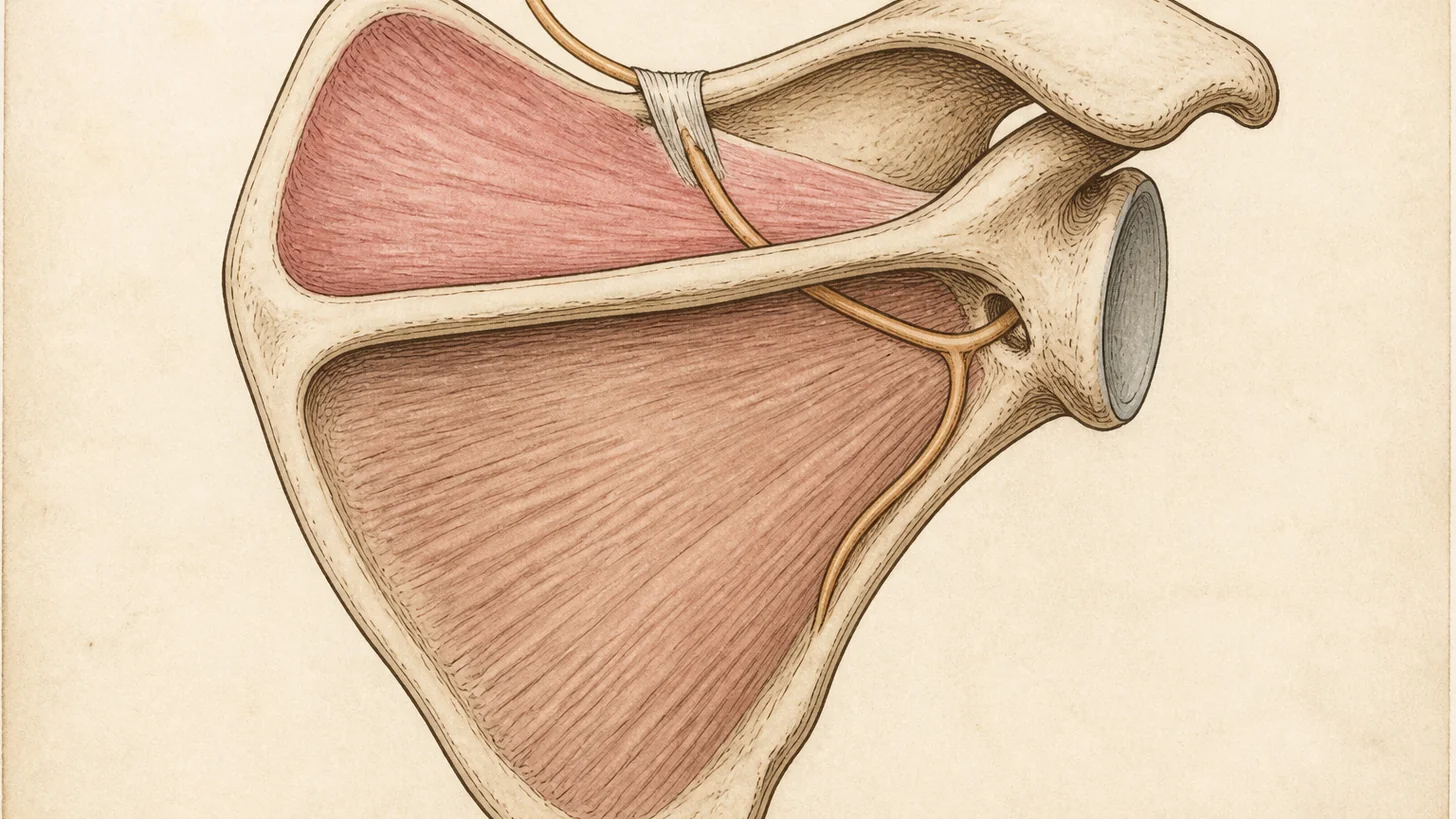
Suprascapular nerve entrapment. The nerve that supplies the supraspinatus and infraspinatus passes through two notches on the shoulder blade, and it can be compressed at either by a thickened ligament, a cyst from a labral tear, or repetitive overhead traction in swimmers, volleyball players, and weightlifters. The pain is a deep, poorly localized ache at the back and top of the shoulder that does not behave like a cuff problem — no painful arc, no relief from a subacromial injection — with weakness lifting the arm out to the side or rotating it outward, and in longer-standing cases visible wasting of the infraspinatus below the spine of the scapula. An ultrasound-guided trial injection of a small volume of local anesthetic at the nerve is the test. If it clearly relieves the usual pain, Dr. Movshis refers to a surgeon who focuses exclusively on peripheral nerve entrapment. The full picture is on our suprascapular nerve entrapment guide.
Referred pain from the neck. A C5 or C6 nerve root irritated by a disc or a bone spur sends pain into the shoulder and the outer arm, and a cervical facet joint refers pain to the top of the shoulder and the inner edge of the shoulder blade. The tells are that the pain travels past the shoulder, that turning or tilting the head reproduces it, that there is tingling or numbness in the hand, and that the shoulder itself examines normally. Dr. Movshis examines the neck on every shoulder patient because this pattern is easy to miss and a shoulder injection will not help it. The treatment is a cervical epidural steroid injection for the nerve root, or a medial branch block and radiofrequency ablation when the facet is the source, both through our neck pain pathway. Our guide to a pinched nerve in the neck covers how to tell.
The joints: AC and glenohumeral arthritis. Acromioclavicular joint pain sits on the very top of the shoulder over the bump where the collarbone meets the shoulder blade, is reproduced by reaching across the chest and by bench press and dips, and is common in weightlifters and after a separation. Glenohumeral osteoarthritis produces a grinding, stiff shoulder in patients usually over 60, and looks like frozen shoulder on exam until the X-ray shows the joint space narrowing. Both can respond to an ultrasound-guided joint injection. The AC joint is small, and in pooled trials landmark-guided injections reached it only 68.2% of the time (Aly 2015). Advanced glenohumeral arthritis is referred for a surgical opinion once injections stop lasting. Our joint pain page covers the arthritis pathway.
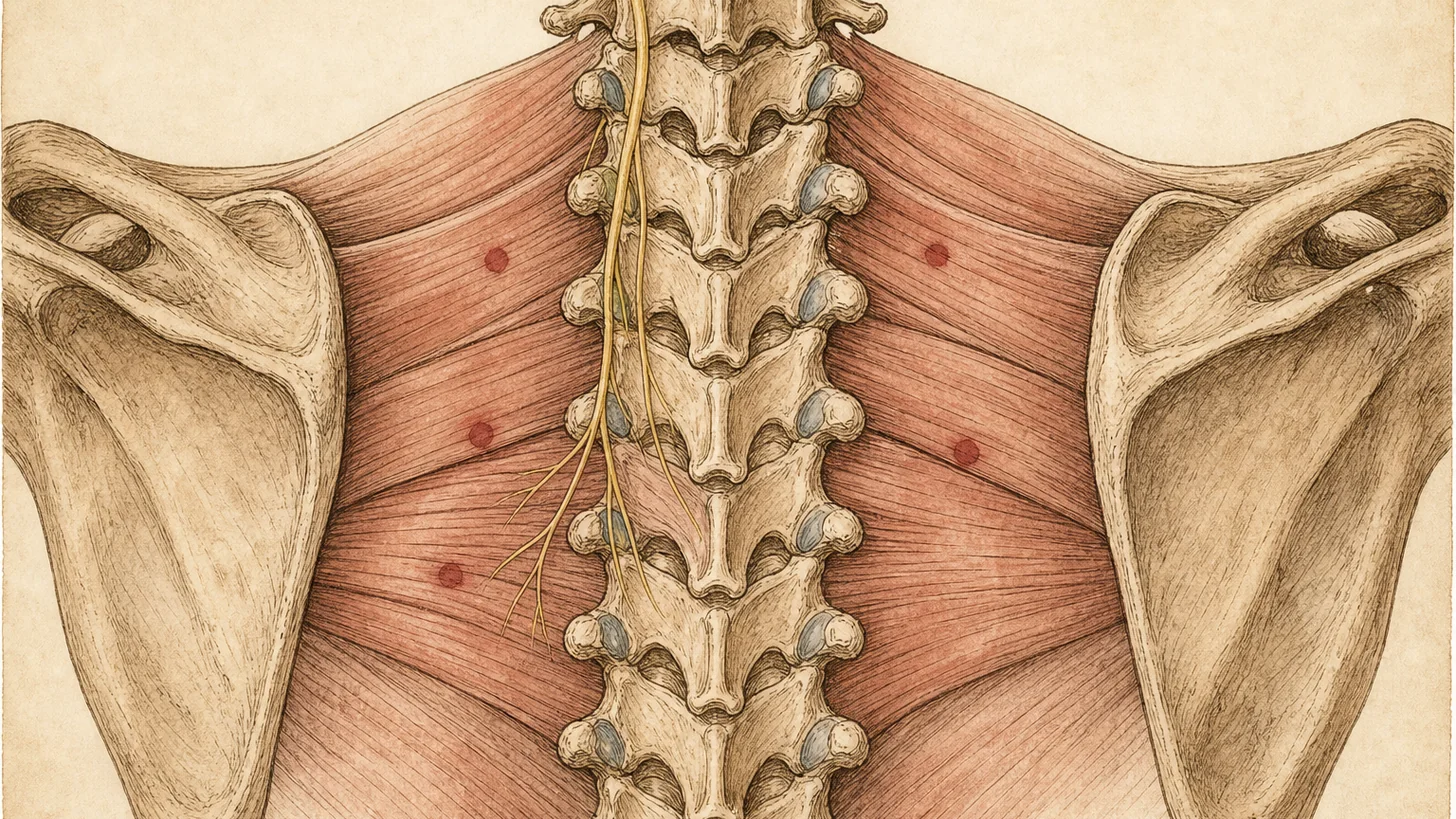
Pain between the shoulder blades belongs to a different list — rhomboid and middle trapezius trigger points, cervical facet referral, a thoracic facet or rib joint — and is covered in our guide to pain between the shoulder blades, with trigger point injections as the usual first procedure when exercise has not settled it.
The red flags that skip the sorting
Left shoulder or arm pain with chest pressure, sweating, shortness of breath, or nausea is a heart problem until proven otherwise. Call 911 or go to the nearest emergency room, not a shoulder appointment. Shoulder pain after a fall or a sudden lifting injury with visible deformity, inability to lift the arm, or immediate marked weakness needs same-day imaging for dislocation, fracture, or an acute full-thickness cuff tear, and a visible deformity means calling 911 or going to the nearest emergency room. A hot, swollen shoulder with fever can be a joint infection: call 911 or go to the nearest emergency room. Night pain that is unrelieved by any position in a smoker or a patient with weight loss, or shoulder pain with a drooping eyelid and a small pupil on the same side, needs a chest X-ray for a tumor at the top of the lung. We see these rarely, and we send them to the right place the same day.
Why the examination and ultrasound settle it
No single shoulder test makes a diagnosis. A large meta-analysis of shoulder examination tests concluded that no single test can be recommended on its own, and that combining tests helps, though only marginally (Hegedus 2012). That is how the sorting above works: the direction of motion loss, the painful arc, the strength tests, the neck maneuvers and the location of tenderness are read together. Ultrasound shows the rotator cuff tendons, the subacromial bursa, the biceps tendon, the AC joint and the suprascapular notch while you move, which MRI cannot do, and it guides the needle. In the trials pooled by Aly and colleagues, ultrasound-guided injections were accurate in 93.6% of AC joint injections versus 68.2% by landmarks (three studies), and in single studies 92.5% versus 72.5% for the shoulder joint and 86.7% versus 26.7% for the biceps sheath (Aly 2015). A shoulder injection that missed looks the same as an injection into the wrong structure, and both end with “cortisone didn’t work.”
MRI has a different job. It is the test for surgical planning, for a suspected full-thickness tear in an active patient, and for the labrum. It is a poor test for “what hurts,” because abnormal findings are the norm in shoulders that do not hurt: in 96 people with no shoulder symptoms, 34% had a rotator cuff tear on MRI, and 54% of those over 60 did (Sher 1995). If you already have an MRI, bring it. It is read alongside the exam.
What gets injected here, and what does not
Corticosteroid is the fast tool. Placed under ultrasound into the subacromial bursa, the glenohumeral joint or the AC joint, it usually eases pain within days and buys weeks to months of function, which is what a bursitis flare, an early frozen shoulder or an arthritic joint needs. It is not injected repeatedly into a tendon, because repeated steroid exposure may weaken tendon tissue, and it is not the answer for a chronic degenerated rotator cuff. PRP is the slow tool, and for the rotator cuff its case is weak: in a placebo-controlled trial of 40 patients, a subacromial PRP injection was no better than saline at one year when both groups exercised (Kesikburun 2013). It is self-pay and offered only after that conversation. Nerve procedures are for the nerve problems: a trial injection of local anesthetic at the suprascapular nerve for suspected entrapment, with surgical referral when it clearly relieves the pain, and a suprascapular nerve block for the night pain of frozen shoulder. Cervical procedures are for the neck: the epidural for the nerve root, the medial branch block and radiofrequency ablation for the facet. Every one is placed under ultrasound or fluoroscopy, takes 10–15 minutes, and you use the arm the same day.
What we do not do is surgery, and knowing when to refer for it is part of the visit. A full-thickness, retracted rotator cuff tear in a patient under 65 who wants to keep using the arm overhead is repaired better early, before the muscle turns to fat and the repair stops holding. Recurrent dislocation, a large labral tear in a young athlete, and glenohumeral arthritis that has stopped responding to injections are surgical problems. We say so at the first visit and send the exam and ultrasound findings with the referral. Physical therapy with Modal’s in-house physical therapists, on the same floor, runs alongside every pathway on this page, and the physical therapy page describes how it works.
What to expect at Modal Pain Management
The first visit is a consultation that takes 45–60 minutes and may include an ultrasound exam. Bring any imaging you have and the list of what has been tried. Dr. Movshis takes the history, examines the shoulder and the neck, orders imaging if the exam calls for it, and tells you which of the five is most likely and why. If an injection is indicated it is typically scheduled within 1–2 weeks at the same office, and physical therapy is arranged to start once the injection has settled the pain enough to load the shoulder. Follow-up at 4–6 weeks measures the response and decides whether the plan holds, escalates to PRP or a nerve procedure, or goes to a surgical opinion. The consultation and image-guided procedures are covered by most commercial PPO plans, some with prior authorization that our office handles. PRP is self-pay and, like every procedure, is priced on the pricing page. The practice does not accept Medicare, Medicaid, HMO plans, or workers’ compensation. You can verify your insurance before the visit.
If your shoulder has had a cortisone shot that did not last, or a physical therapy course that did not change anything, it is worth checking whether the right structure was identified. That is the visit to book.
References
- Hegedus EJ, Goode AP, Cook CE, et al. Which physical examination tests provide clinicians with the most value when examining the shoulder? Update of a systematic review with meta-analysis of individual tests. Br J Sports Med. 2012;46(14):964–978. doi:10.1136/bjsports-2012-091066 · PubMed
- Aly AR, Rajasekaran S, Ashworth N. Ultrasound-guided shoulder girdle injections are more accurate and more effective than landmark-guided injections: a systematic review and meta-analysis. Br J Sports Med. 2015;49(16):1042–1049. doi:10.1136/bjsports-2014-093573 · PubMed
- Kelley MJ, Shaffer MA, Kuhn JE, et al. Shoulder pain and mobility deficits: adhesive capsulitis. Clinical practice guidelines linked to the International Classification of Functioning, Disability, and Health from the Orthopaedic Section of the American Physical Therapy Association. J Orthop Sports Phys Ther. 2013;43(5):A1–A31. doi:10.2519/jospt.2013.0302 · PubMed
- Kesikburun S, Tan AK, Yilmaz B, Yaşar E, Yazicioğlu K. Platelet-rich plasma injections in the treatment of chronic rotator cuff tendinopathy: a randomized controlled trial with 1-year follow-up. Am J Sports Med. 2013;41(11):2609–16. doi:10.1177/0363546513496542 · PubMed
- Kuhn JE, Dunn WR, Sanders R, et al. Effectiveness of physical therapy in treating atraumatic full-thickness rotator cuff tears: a multicenter prospective cohort study. J Shoulder Elbow Surg. 2013;22(10):1371–1379. doi:10.1016/j.jse.2013.01.026 · PubMed
- Sher JS, Uribe JW, Posada A, Murphy BJ, Zlatkin MB. Abnormal findings on magnetic resonance images of asymptomatic shoulders. J Bone Joint Surg Am. 1995;77(1):10–15. doi:10.2106/00004623-199501000-00002 · PubMed
- Zreik NH, Malik RA, Charalambous CP. Adhesive capsulitis of the shoulder and diabetes: a meta-analysis of prevalence. Muscles Ligaments Tendons J. 2016;6(1):26–34. doi:10.11138/mltj/2016.6.1.026 · PubMed
- Challoumas D, Biddle M, McLean M, Millar NL. Comparison of treatments for frozen shoulder: a systematic review and meta-analysis. JAMA Netw Open. 2020;3(12):e2029581. doi:10.1001/jamanetworkopen.2020.29581 · PubMed