Runners can usually name the mile marker. Two miles in, sometimes four, a sharp ache lands on the outside of the knee just above the joint. Walk for a minute and it fades. Start running again and it is back within a few hundred meters, worse than before, and a long downhill turns it into a stabbing pain that forces a straight-legged shuffle. By the next morning the knee feels almost normal, which is why so many people keep trying to run through it.
That is iliotibial band syndrome (ITBS), the most common cause of pain on the outside of the knee in runners, with an estimated incidence between 5% and 14% (van der Worp 2012). In a clinic series of 2,002 running injuries it was second only to patellofemoral pain (Taunton 2002). Jumps in training load are among the most consistent risk factors for knee injuries in runners (Senthil 2026), which usually means the weeks of peak mileage before a goal race.
Why it hurts: compression of a sensitive fat layer, not a band rubbing back and forth
For decades the explanation was friction. The band was said to flick forward and back over the bony prominence on the outside of the femur, the lateral femoral epicondyle, thousands of times per run, until a bursa beneath it became inflamed. The older literature calls the condition iliotibial band friction syndrome for that reason.
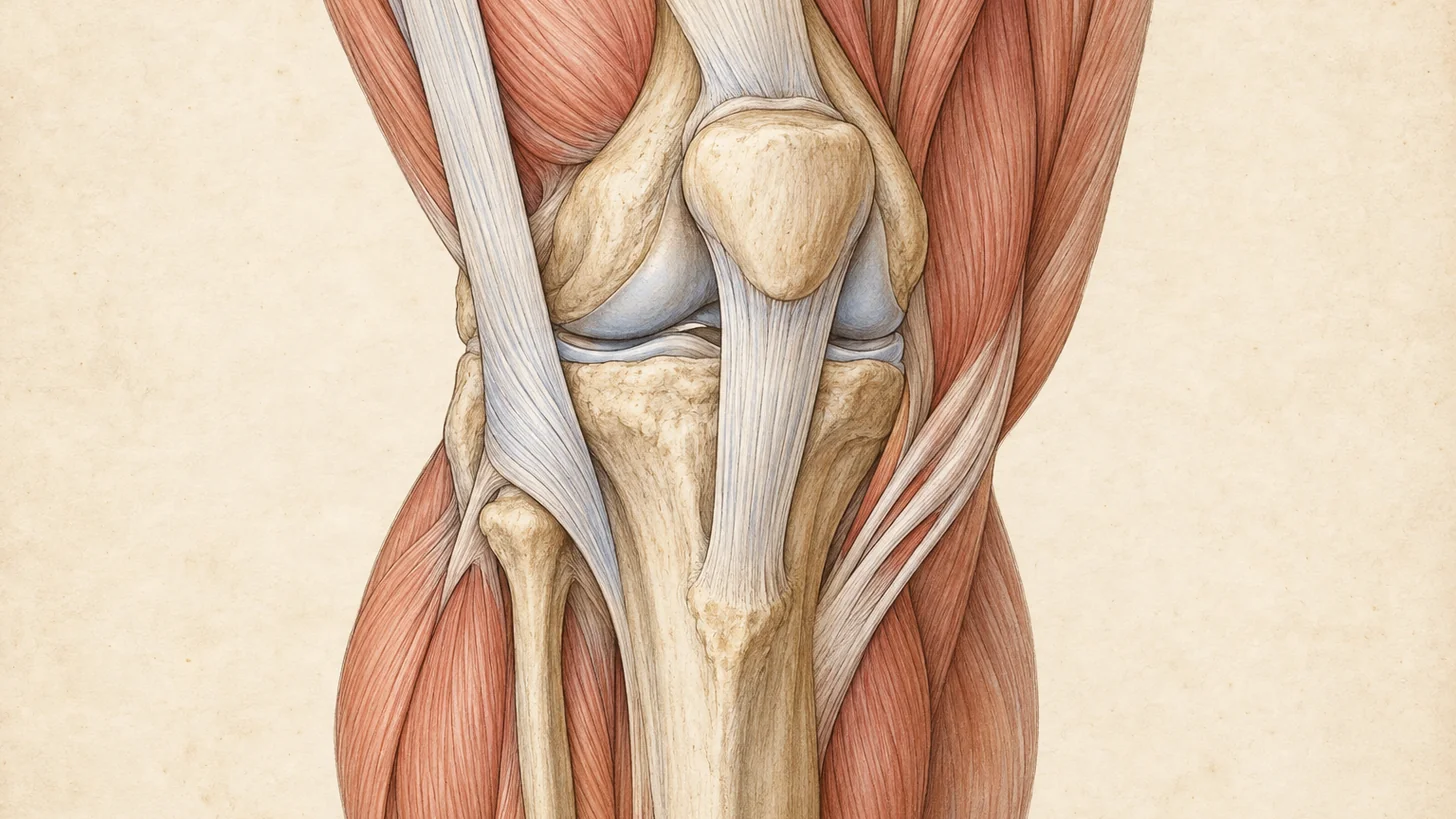
Fairclough and colleagues then dissected 15 cadavers and imaged healthy volunteers and athletes with the syndrome. In every specimen the band was anchored to the lower femur by fibrous strands and sat on a layer of richly innervated, vascular fat. No bursa appeared in any cadaver, volunteer, or patient. MRI showed the band pressed against the epicondyle at about 30 degrees of knee flexion, driven by inward rotation of the tibia, and in the two injured athletes the abnormal signal sat in that fat layer, not in the band (Fairclough 2006). Their follow-up paper was blunt: the band cannot roll over the epicondyle at all, the impression of movement is an illusion created by shifting tension in its front and back fibers, and the pain comes from compression of the fat and loose connective tissue between band and bone (Fairclough 2007). A separate cadaver study found the band firmly attached along the femur from the greater trochanter down to and including the lateral condyle (Falvey 2010).
Running biomechanics fit that picture. In runners with the syndrome, the painful contact occurred near footstrike with the knee bent an average of 21.4 degrees, at or just below the 30 degrees classically described (Orchard 1996). Downhill running lands the knee straighter, right inside that zone, which is why a long downhill is where the pain peaks. The same study found that faster running and sprinting were less provocative, because the knee is already bent past the zone at footstrike (Orchard 1996). Noble, who first described the condition in runners, noted the pain may be prevented by walking with a stiff knee (Noble 1980).
The mechanism also defines the injection target. If the pain generator is the innervated fat between the band and the epicondyle, then that layer, not “the band” in the abstract, is where medication belongs, and that is where it is placed under ultrasound.
What presses the band into the bone
Prospective gait data point upstream, to the hip. Female recreational runners underwent gait analysis and were followed for two years, and the 18 who developed ITBS had shown greater peak hip adduction and knee internal rotation at baseline than matched controls (Noehren 2007). In plain terms, the thigh drifts inward and rotates as the foot lands, which tensions the band and drives it into the epicondyle.
Hip abductor strength belongs to the same story. In Fredericson’s clinic series, injured runners were weaker in hip abduction on the painful side than on the other side and than healthy controls (Fredericson 2000), and a 2023 meta-analysis found lower isometric abductor strength in female runners with current ITBS (Foch 2023). The evidence is not unanimous. An earlier systematic review judged the role of abductor weakness unclear (van der Worp 2012), and some of the weakness may follow the pain rather than cause it. That does not change the plan, because the hip gets strengthened either way.
The runner’s story that makes the diagnosis
ITBS is diagnosed from the history and on the exam table. The story is pain on the outside of the knee, a thumb’s width or two above the joint line, that starts at a reproducible distance or time into a run and builds if you keep going. It is worse downhill and going down stairs, quiet at rest, and the knee does not swell, lock, or give way. Many runners first notice it after a jump in weekly mileage, a new hilly route, or a block of track sessions run in one direction. A treatment review by Fredericson lists exactly those training errors: excessive running in the same direction on a track, higher than normal weekly mileage, and downhill running (Fredericson 2005).
Two exam findings carry most of the weight.
The Noble compression test. Lying on your back with the knee bent to 90 degrees, Dr. Movshis presses a thumb over the lateral femoral epicondyle and slowly straightens the knee. Reproduction of your running pain at roughly 30 degrees of flexion is a positive test, and it maps onto the compression zone described above.
The Ober test. Lying on the good side, the top leg is brought back and allowed to drop toward the table. If it stays up, the hip lacks adduction range. Read it as a test of the hip and the tensor fasciae latae rather than of band length, because the band itself is fixed to the femur and lengthens by less than half a percent under load (Falvey 2010).
The rest of the exam looks for what the knee is not telling you: single-leg squat and step-down control, abductor strength side to side, the lumbar spine and nerve tension signs, the fibular head, and the lateral joint line. Bedside ultrasound at the same visit shows the band and the tissue beneath it at the epicondyle, the lateral collateral ligament, the biceps femoris tendon, and the outer rim of the lateral meniscus. MRI is not needed to make the diagnosis. It is ordered when the story or exam points inside the joint or at bone.
When outside-knee pain is not the IT band
Several neighbors of the band get called “IT band” by the time a runner reaches us. The decision rules we use:
- Lateral meniscus tear. If the pain started with a twist or pivot, sits right on the joint line, and comes with clicking, catching, locking, or swelling, think meniscus. That needs MRI and often an orthopedic opinion before any return to running, arranged through our sports medicine pathway.
- Lateral collateral ligament sprain. Usually follows a blow or an outward-bending force on the knee. The tenderness is on the cord-like ligament running from the epicondyle to the head of the fibula, and stressing the knee outward reproduces it.
- Patellofemoral pain. Around or behind the kneecap rather than on the side, worse on stairs, squats, and after long sitting. It is the most common running knee injury (Taunton 2002) and has its own plan, laid out in knee pain after running and built around physical therapy.
- Biceps femoris tendinopathy. Back-and-outside of the knee near the fibular head, provoked by resisted knee bending, hill sprints, and fast finishes.
- Proximal tibiofibular joint. Pain right at the top of the fibula, sometimes with a sense of shifting, sometimes stirred up by ankle movement.
- Common peroneal nerve. Burning or numbness on the outer shin and top of the foot, weakness lifting the foot or toes, and tingling when the nerve is tapped where it wraps the fibular neck. That is a nerve problem, covered on our peripheral nerve entrapment page.
- Lumbar radiculopathy. Pain that travels from the low back or buttock down the outside of the leg, with numbness or weakness and a positive straight-leg raise, is usually an L5 nerve root. The spine gets treated, see radiculopathy.
- Proximal IT band pain and GTPS. Pain at the outside of the hip, worst lying on that side, is usually gluteal tendinopathy at the greater trochanter rather than the band. See hip bursitis.
Two more belong on the list in specific runners. Over 50, with morning stiffness and aching after rather than during runs, lateral compartment arthritis is possible and the knee osteoarthritis workup applies. And pain at rest or at night, pain hopping on the leg, or tenderness over bone rather than soft tissue raises a bone stress injury, which means MRI before another run.
What treats IT band syndrome, in the order we use it
A systematic review of 24 studies and 1,012 patients found improvement reported in every treatment category, and concluded that most of them cannot yet be recommended over one another because diagnosis and methods vary so much between studies. The authors recommended structured rehabilitation as the reasonable first step (Ferrero 2026). That matches what we do.
Change the load instead of stopping everything
Complete rest deconditions the hip that needs training. The better rule: if pain starts at mile four, run three on flat ground and stop while the knee is quiet. Cut downhill running, heavily cambered roads, and laps in one direction on a track. Faster strides are often tolerated better than slow jogging early on, which follows from the knee angles at footstrike (Orchard 1996) and is part of Fredericson’s return protocol (Fredericson 2005). Cyclists get ITBS too (Fairclough 2006), so if a stationary bike reproduces the pain, swap it for pool running or swimming while the knee settles.
Strengthen the hip
This is where the most consistent signal sits. In Fredericson’s series, 24 runners with ITBS completed a six-week program aimed at the gluteus medius. Abductor torque rose 34.9% in the women and 51.4% in the men, 22 of 24 were pain free and back to running at six weeks, and none had recurred at six months (Fredericson 2000). There was no control group, so some of that recovery would have happened anyway. A 2024 systematic review of 13 studies and 201 runners found hip abductor strengthening to be a common element across the programs, with pain reductions ranging from 27% to 100% over two to eight weeks, although the studies were too different to pool (Sanchez-Alvarado 2024).
Our in-house physical therapy program starts with side-lying abduction and side-plank work, moves to single-leg bridges, step-downs, and lateral band walks, and finishes with single-leg strength and hopping drills that look like running. Fredericson’s review recommends emphasizing eccentric control and integrated, multi-plane movement (Fredericson 2005), and that is the progression we use.
Retrain the stride
A small increase in step rate changes how the knee and hip take load. In 45 healthy recreational runners, raising cadence 5% or 10% above preferred reduced energy absorbed at the knee, and the 10% increase also reduced peak hip adduction angle along with the hip adduction and internal rotation moments (Heiderscheit 2011). Those are the same motions that predicted ITBS in Noehren’s cohort. The caveat is real: the study was in healthy runners, and gait retraining for ITBS itself has not been tested in rigorous trials (Sanchez-Alvarado 2024). In practice, a metronome app set about 5% above your usual cadence on easy runs is cheap and low-risk.
Why rolling and stretching the band does so little
The band is a thickened part of a sleeve of fascia that wraps the thigh and is fixed to the femur along most of its length. Falvey’s group measured strain in cadaver bands during three standard stretches and movement of the band in living volunteers, and mean band lengthening was under 0.5% (Falvey 2010). In a randomized trial of 30 adults, a session of foam rolling or stretching produced no measurable change in band stiffness on shear-wave elastography (Pepper 2021). In a separate crossover study, rolling the gluteal muscles improved hip adduction range by 14.8%, while rolling the band itself improved it by 2% (Hall 2018).
A balanced review found some support for stretching in early rehabilitation and no direct evidence of harm (Opara 2023). So if rolling or stretching makes you feel better before a run, there is no reason to forbid it. Aim it at the tensor fasciae latae and the glutes, keep the pressure off the sore spot at the outer knee, and do not let it replace the strength work.
Ultrasound-guided corticosteroid injection
The best trial here is small. Gunter and Schwellnus randomized 18 runners with ITBS of less than two weeks’ duration to methylprednisolone 40 mg plus local anesthetic or local anesthetic alone, injected where the band crosses the lateral femoral condyle, then repeated a treadmill running test at day 7 and day 14. Steroid showed a trend toward lower running pain by day 7 (p = 0.07) and a significant reduction between day 7 and day 14 (Gunter 2004). That is real but short-horizon evidence in recent-onset cases, with landmark rather than image-guided injections. Older and uncontrolled, Noble’s series of 100 knees found that only 30 of the 73 followed up settled on a single injection plus reduced training (Noble 1980).
At Modal Pain Management, Dr. Movshis places the injection under ultrasound into the fat and connective tissue beneath the band at the lateral femoral epicondyle, the layer Fairclough identified as the pain source. The point is a window of pain-free training in which the hip work can happen, and it is not repeated as a substitute for that work. The procedure itself is described on the joint and soft tissue injections page.
PRP for refractory cases
Platelet-rich plasma is offered for runners whose pain has persisted through at least three months of proper rehabilitation. The ITBS-specific evidence is thin. The 2026 systematic review found one adequately powered placebo-controlled trial of ultrasound-guided PRP in refractory ITBS with a favorable result, and flagged that it has not been independently replicated (Ferrero 2026). PRP takes weeks to act and often leaves the area sore for several days, so it is never a pre-race option.
When we refer
Percutaneous tenotomy is not something we offer, and we refer for it. Surgical release or lengthening of the back part of the band at the epicondyle is reserved for the rare runner who fails all of the above. A 2020 systematic review found return-to-sport rates of 81% to 100% in the surgical studies that reported it, with most of that evidence coming from low-level case series (Bolia 2020).
If your race is a few weeks away
If the knee is acting up in the weeks before a marathon or half marathon, these are the decision rules we use with runners in the office.
If pain starts late in long runs and walking is normal, keep the taper. Do the abductor work three or four days a week, raise cadence slightly, and cut downhill running from every remaining session.
If pain starts in the first few miles, have an honest conversation with yourself. In a marathon, pain at mile four means 22 miles of altered mechanics, and the compensation injuries that follow, in the hip, the other knee, or the foot, can outlast the IT band problem. Sitting this one out is a legitimate medical decision.
If you are thinking about an injection, it is considered only for recent-onset pain, and only with enough lead time to test the knee on several runs before race day. The trial evidence covers two weeks of treadmill running after an injection given within two weeks of symptom onset (Gunter 2004), not a marathon. It is never a way to mask pain for race day, and an injection in the last few days risks post-injection soreness on the start line.
On race day, use a run-walk pattern from the first mile rather than waiting for pain to force it. Shorten your stride and quicken your cadence on every downhill section of the course, and most of all on the long descents. Nothing new on race day, including shoes, insoles, or a knee strap you have never run in. And if you are limping, stop.
Returning to running after ITBS
We clear a runner to start back when they can walk briskly and take stairs down without pain, the Noble compression test no longer reproduces the running pain, and a slow single-leg squat on the injured side looks like the other side. Then:
- Start with short runs on flat ground every other day, ending well before the old pain point.
- Use quicker strides rather than a slow shuffle in the first weeks (Fredericson 2005).
- Add distance and frequency before adding hills, and add hills before adding downhill repeats.
- Keep the hip strengthening going twice a week after you are back to full mileage, since it is the part most runners drop first.
Fredericson’s protocol expects most runners to recover fully by about six weeks with this approach (Fredericson 2005). A flare during the progression means dropping back to the last pain-free week, not starting over.
Seeing Dr. Movshis for IT band pain
The first visit is a focused history, the exam described above, and bedside ultrasound of the outer knee, usually with a diagnosis and plan the same day. Dr. Movshis is dual board-certified in Anesthesiology and Pain Medicine and completed an ACGME-accredited interventional pain medicine fellowship at the Icahn School of Medicine at Mount Sinai (NPI 1942741160). Injections are placed by him under ultrasound at 369 Lexington Avenue, Floor 25, and physical therapy runs in the same office, so the injection and the strength program are planned together.
Same-week appointments are available. You can verify your insurance before the visit.
If the pain shows up at the same point in every run and three weeks of backing off has not changed that, the exam is the next step, not another foam roller.
References
- van der Worp MP, van der Horst N, de Wijer A, Backx FJ, Nijhuis-van der Sanden MW. Iliotibial band syndrome in runners: a systematic review. Sports Med. 2012;42(11):969–92. doi:10.2165/11635400-000000000-00000 · PubMed
- Taunton JE, Ryan MB, Clement DB, McKenzie DC, Lloyd-Smith DR, Zumbo BD. A retrospective case-control analysis of 2002 running injuries. Br J Sports Med. 2002;36(2):95–101. doi:10.1136/bjsm.36.2.95 · PubMed
- Senthil K, Whitaker S, Nazarov M, et al. Common Risk Factors for Knee Injuries in Runners: A Systematic Review. Orthop J Sports Med. 2026;14(5):23259671261444314. doi:10.1177/23259671261444314 · PubMed
- Fairclough J, Hayashi K, Toumi H, et al. The functional anatomy of the iliotibial band during flexion and extension of the knee: implications for understanding iliotibial band syndrome. J Anat. 2006;208(3):309–16. doi:10.1111/j.1469-7580.2006.00531.x · PubMed
- Fairclough J, Hayashi K, Toumi H, et al. Is iliotibial band syndrome really a friction syndrome? J Sci Med Sport. 2007;10(2):74–6. doi:10.1016/j.jsams.2006.05.017 · PubMed
- Orchard JW, Fricker PA, Abud AT, Mason BR. Biomechanics of iliotibial band friction syndrome in runners. Am J Sports Med. 1996;24(3):375–9. doi:10.1177/036354659602400321 · PubMed
- Noble CA. Iliotibial band friction syndrome in runners. Am J Sports Med. 1980;8(4):232–4. doi:10.1177/036354658000800403 · PubMed
- Noehren B, Davis I, Hamill J. ASB clinical biomechanics award winner 2006 prospective study of the biomechanical factors associated with iliotibial band syndrome. Clin Biomech (Bristol). 2007;22(9):951–6. doi:10.1016/j.clinbiomech.2007.07.001 · PubMed
- Fredericson M, Cookingham CL, Chaudhari AM, Dowdell BC, Oestreicher N, Sahrmann SA. Hip abductor weakness in distance runners with iliotibial band syndrome. Clin J Sport Med. 2000;10(3):169–75. doi:10.1097/00042752-200007000-00004 · PubMed
- Foch E, Brindle RA, Pohl MB. Lower extremity kinematics during running and hip abductor strength in iliotibial band syndrome: A systematic review and meta-analysis. Gait Posture. 2023;101:73–81. doi:10.1016/j.gaitpost.2023.02.001 · PubMed
- Fredericson M, Wolf C. Iliotibial band syndrome in runners: innovations in treatment. Sports Med. 2005;35(5):451–9. doi:10.2165/00007256-200535050-00006 · PubMed
- Falvey EC, Clark RA, Franklyn-Miller A, Bryant AL, Briggs C, McCrory PR. Iliotibial band syndrome: an examination of the evidence behind a number of treatment options. Scand J Med Sci Sports. 2010;20(4):580–7. doi:10.1111/j.1600-0838.2009.00968.x · PubMed
- Ferrero A, Moro A, Minelli M, et al. Extreme Diagnostic and Therapeutic Heterogeneity Limits Treatment Conclusions in Iliotibial Band Syndrome: A Systematic Review with Level-of-Evidence Stratification. Orthop Res Rev. 2026;18:629195. doi:10.2147/ORR.S629195 · PubMed
- Sanchez-Alvarado A, Bokil C, Cassel M, Engel T. Effects of conservative treatment strategies for iliotibial band syndrome on pain and function in runners: a systematic review. Front Sports Act Living. 2024;6:1386456. doi:10.3389/fspor.2024.1386456 · PubMed
- Heiderscheit BC, Chumanov ES, Michalski MP, Wille CM, Ryan MB. Effects of step rate manipulation on joint mechanics during running. Med Sci Sports Exerc. 2011;43(2):296–302. doi:10.1249/MSS.0b013e3181ebedf4 · PubMed
- Pepper TM, Brismée JM, Sizer PS, et al. The Immediate Effects of Foam Rolling and Stretching on Iliotibial Band Stiffness: A Randomized Controlled Trial. Int J Sports Phys Ther. 2021;16(3):651–661. doi:10.26603/001c.23606 · PubMed
- Hall M, Chadwick Smith J. The effects of an acute bout of foam rolling on hip range of motion on different tissues. Int J Sports Phys Ther. 2018;13(4):652–660. PubMed
- Opara M, Kozinc Ž. Stretching and Releasing of Iliotibial Band Complex in Patients with Iliotibial Band Syndrome: A Narrative Review. J Funct Morphol Kinesiol. 2023;8(2):74. doi:10.3390/jfmk8020074 · PubMed
- Gunter P, Schwellnus MP. Local corticosteroid injection in iliotibial band friction syndrome in runners: a randomised controlled trial. Br J Sports Med. 2004;38(3):269–72. doi:10.1136/bjsm.2003.000283 · PubMed
- Bolia IK, Gammons P, Scholten DJ, Weber AE, Waterman BR. Operative Versus Nonoperative Management of Distal Iliotibial Band Syndrome-Where Do We Stand? A Systematic Review. Arthrosc Sports Med Rehabil. 2020;2(4):e399–e415. doi:10.1016/j.asmr.2020.04.001 · PubMed
- Brosseau L, Casimiro L, Milne S, et al. Deep transverse friction massage for treating tendinitis. Cochrane Database Syst Rev. 2002;(4):CD003528. doi:10.1002/14651858.CD003528 · PubMed