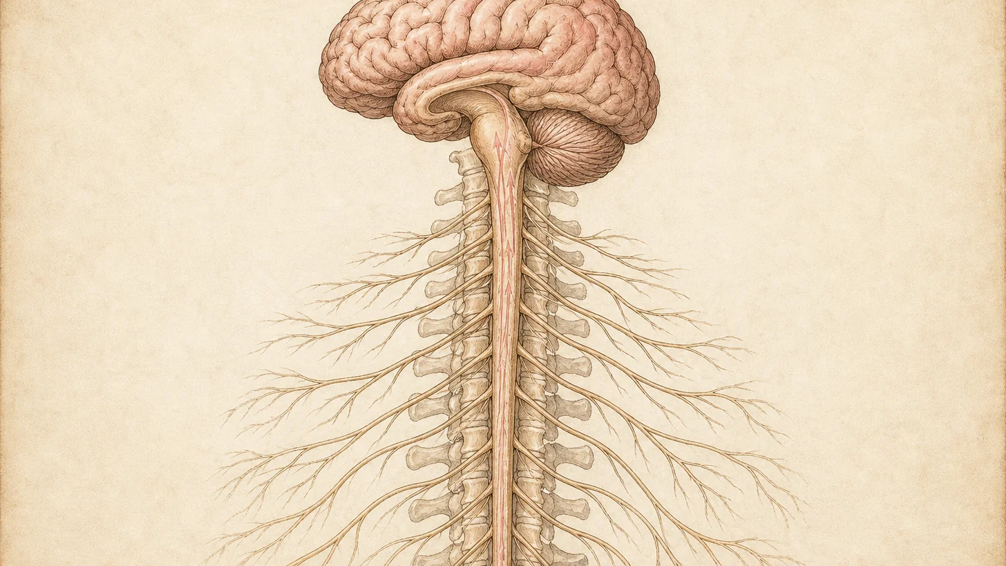
Neck pain affects millions of people worldwide and can significantly impact your daily life, affecting your ability to work, exercise, and enjoy activities you love. Whether you’re experiencing sharp, shooting pain or a constant dull ache, neck discomfort can be both physically limiting and emotionally frustrating. At Modal Pain Management NYC, located at 369 Lexington Avenue in Manhattan, Dr. Alex Movshis and our team specialize in advanced non-surgical interventional pain management techniques that address the root cause of your neck pain. Our approach focuses on providing effective relief without the risks associated with surgery, allowing you to return to the activities that matter most to you. The cervical spine is delicate and complex, containing nerves, discs, and joints that can become injured or irritated. Accurate diagnosis is essential for safe and effective treatment, which is why Dr. Movshis takes the time to thoroughly evaluate each patient and develop a personalized treatment plan.
Common Causes of Neck Pain
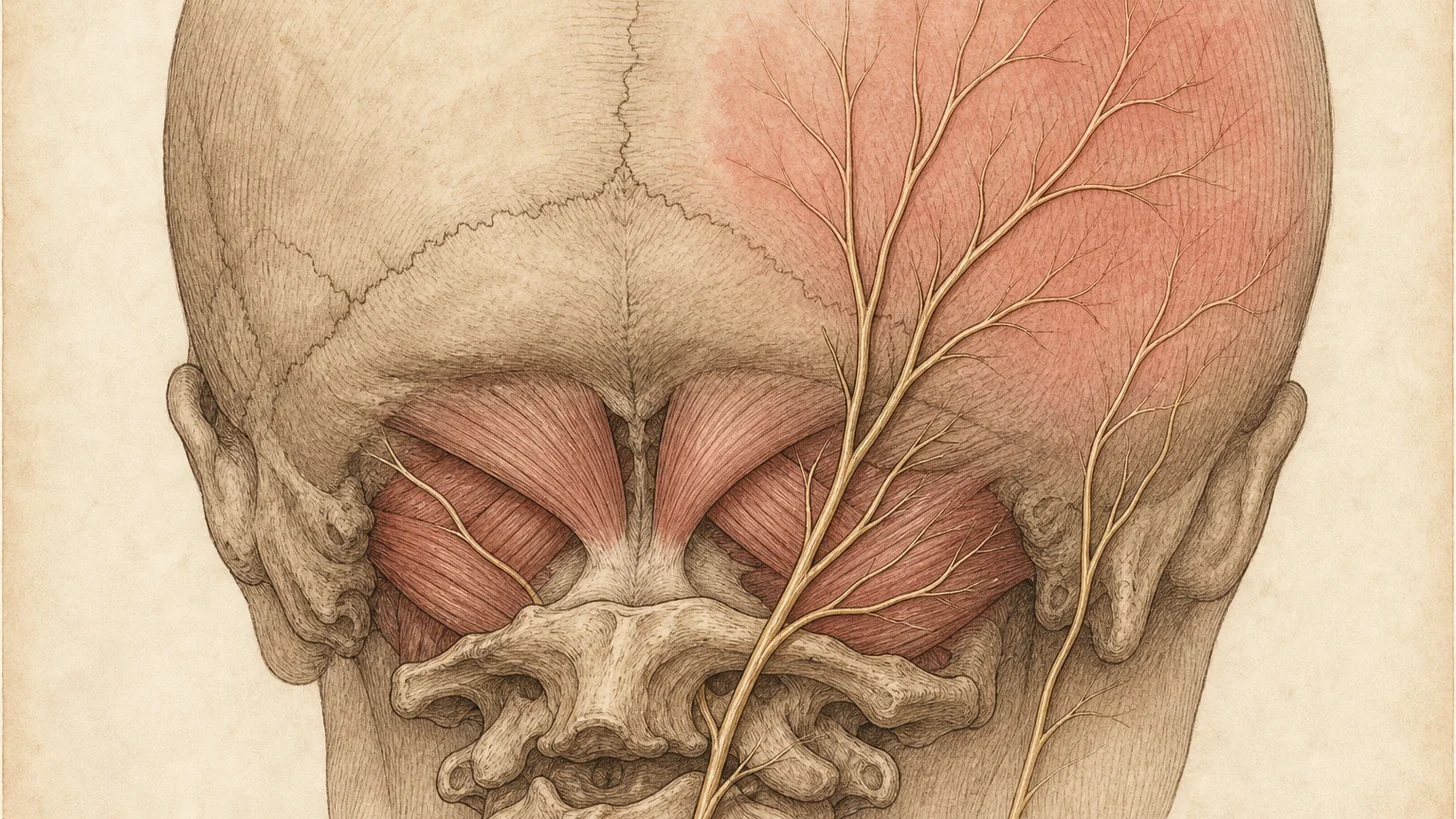
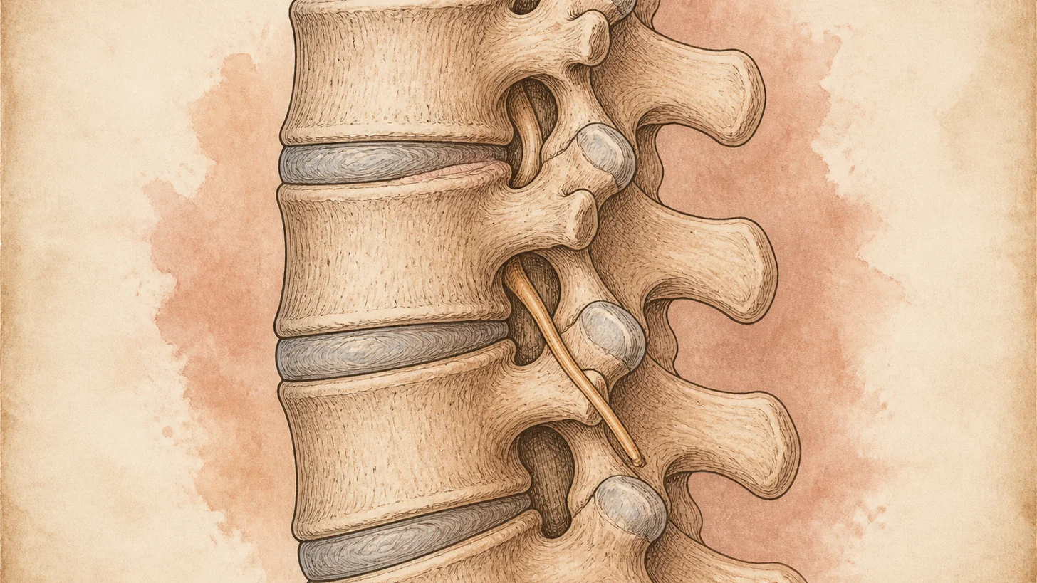
Neck pain can originate from various structures in the cervical spine, each requiring a specific treatment approach. Cervical disc herniation occurs when the gel-like material inside a spinal disc ruptures and irritates nearby nerves, often causing radiating pain down the arm. Degenerative disc disease involves the breakdown of spinal discs over time, leading to chronic pain and stiffness. Whiplash injuries, commonly associated with car accidents, can cause acute and delayed neck pain through sudden forceful movement. Cervical radiculopathy develops when a nerve root in the neck becomes pinched or compressed, resulting in sharp pain, numbness, or tingling in the arms and hands. Muscle strain from poor posture, repetitive motions, or sudden trauma can cause localized neck pain and tension. Cervical facet syndrome occurs when the small joints at the back of the spine become arthritic or irritated, contributing to neck stiffness and pain. Our team carefully evaluates each patient to identify which conditions are contributing to your specific symptoms. For a comprehensive overview of multiple neck pain causes, see 10 common causes of chronic neck pain.
When to Seek Treatment
If you’re experiencing neck pain that persists for more than a few days or is severe enough to interfere with your daily activities, it’s time to seek professional evaluation. You should also consider treatment if you have numbness or tingling radiating into your arms or hands, if over-the-counter medications haven’t provided relief, or if your pain is limiting your ability to work or exercise. Some warning signs that warrant immediate attention include severe pain following trauma, weakness in your arms or hands, or difficulty with coordination. Early intervention often leads to better outcomes, as we can identify and address the underlying cause before the condition progresses. Dr. Movshis and the clinic team are here to provide compassionate, expert care that gets you back to feeling your best.
Diagnosis and Assessment
We begin with a comprehensive evaluation that includes a detailed medical history, physical examination, and if necessary, advanced imaging such as MRI or CT scans. These diagnostic tools help us visualize the cervical spine and identify exactly where your pain is originating. We assess your range of motion, reflexes, and neurological function to understand how your condition is affecting you. We may also perform provocative testing to pinpoint which specific joints or nerve roots are involved in your pain. This thorough assessment ensures that we develop a treatment plan targeted specifically at your condition, maximizing the chances of successful relief and long-term improvement.
Treatment Options
We utilize a range of advanced minimally invasive procedures to provide targeted relief for neck pain. Cervical epidural steroid injections deliver medication directly to the inflamed tissues surrounding the spinal nerves, reducing inflammation and pain signals. Cervical facet joint injections target the small joints at the back of the spine where arthritis and inflammation often develop. Cervical nerve blocks interrupt pain signals traveling along specific nerves, providing relief while allowing you to participate in physical therapy. Trigger point injections address muscle tension and pain by injecting medication directly into affected muscles. We also emphasize physical therapy and rehabilitation as essential components of treatment, helping to strengthen supporting muscles and improve flexibility. Each procedure is performed under fluoroscopic guidance with ultrasound or X-ray visualization to ensure precision and safety. Most patients experience significant improvement with our comprehensive treatment approach, which combines procedures with rehabilitative care.
What to Expect
When you visit our clinic for neck pain treatment, you can expect a professional, compassionate experience focused on your comfort and recovery. Your first appointment will include a thorough consultation and examination, during which Dr. Movshis will discuss your symptoms, review any imaging, and explain treatment options in clear, understandable language. If a procedure is recommended, we’ll schedule it at your convenience and provide detailed pre-procedure instructions. During the procedure itself, we use advanced imaging technology to guide our injections with precision, minimizing risk and maximizing effectiveness. Most procedures take 15 to 30 minutes, and you can typically return home the same day. You may experience mild soreness at the injection site for a day or two, but this is normal and manageable.
Recovery and Prevention
Recovery from neck pain treatment varies depending on your specific condition and the procedures performed. Many patients notice improvement within a few days to a few weeks, with continued improvement over the following weeks as inflammation decreases and muscles strengthen through physical therapy. To prevent future episodes of neck pain, we recommend maintaining good posture, taking regular breaks if you spend extended time at a desk, and performing gentle stretching and strengthening exercises. Ergonomic adjustments to your workspace can make a significant difference in preventing recurrent pain. If you’ve experienced whiplash or trauma, protective techniques can help prevent further injury. Our team provides ongoing guidance and support to help you maintain your pain relief and prevent relapse, ensuring long-term success in managing your cervical spine health.